- 12 year old mn Shih Tzu with iCa=1.7 and PTH in the high normal range. ACTH stim pre=2.2mcg/dL, post=22.6mcg/dL. normal SDMA=10mcg/dL, chems wnl except for Ca=13.0, Na=153, Chol=269. CBC showed thrombocytosis.
- Abdominal US was wnl except for bilateral loss of renal CM definition.
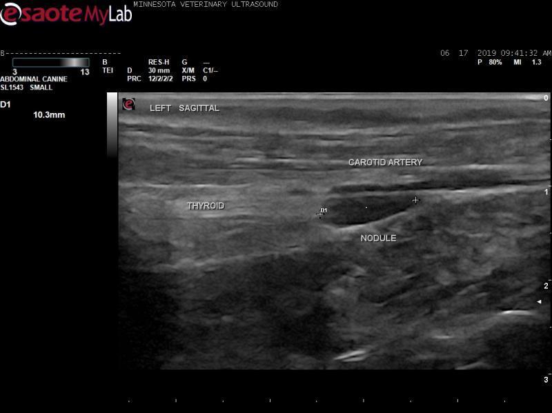
- Cervical ultrasound shows an irregularly shaped hypoechoic nodule in the left thyroid gland and a small hypoechoic nodule in the right thyroid gland. Both nodules were negative for blood flow on Color flow Doppler.
- 12 year old mn Shih Tzu with iCa=1.7 and PTH in the high normal range. ACTH stim pre=2.2mcg/dL, post=22.6mcg/dL. normal SDMA=10mcg/dL, chems wnl except for Ca=13.0, Na=153, Chol=269. CBC showed thrombocytosis.
- Abdominal US was wnl except for bilateral loss of renal CM definition.
- Cervical ultrasound shows an irregularly shaped hypoechoic nodule in the left thyroid gland and a small hypoechoic nodule in the right thyroid gland. Both nodules were negative for blood flow on Color flow Doppler.
- Based upon labwork values and ultrasound findings, primary differential diagnoses are parathyroid adenoma, parathyroid hyperplasia, and parathyroid carcinoma.
- My concern is that this dog had multiple sedative drugs on board (gabapentin, trazadone, clomicalm, butorphanol) and was given IV propofol for this study. Vessels appeared more prominent than normal. I want to be sure that I am not misinterpreting vessels as nodules, especially on the right side. Also, previous parathyroid adenomas I have seen were fairly round in shape. The one on the left side of this dog is elongated.
![]()
![]()
![]()
![]()
4 responses to “12 yr old mn sedated Shih Tzu with hypercalcemia”
Would go for nodules rather
Would go for nodules rather than blood vessels as there is blood flow in the surrounding vessels. Clinical picture fits functional parathyroid nodule.
I’m not convinced that
I’m not convinced that hypoechoic structure adjacent to the carotid is a pth adenoma… unless ectopic but tough to tell where you are there. The third image looks more like a small adenoma or larger parathyroid.
Here are some pth adenomas from the archive for reference
https://sonopath.com/members/case-studies/search?text=parathyroid&species=All
Thank you for your input.
Thank you for your input. EL, which side (R or L), clip are you referring to?
I take that back on tbhe
I take that back on tbhe third image i see that corresponds to your first clip and im seeing the vessel there… lesson never make dx on a still 🙂
if you can get a needle (25g) in that lesion in still image 2 that would be my next step just have to avoid the vessel. Corkscrew technique applies here.