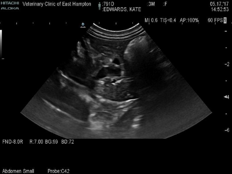
These are images and clips from an abdominal ultrasound on Kate Edwards, a 3 month old mini English Bulldog. She presented for inappropriate urination. U/a revealed 21-50 Ammonium Biurate Crystals per HPF, u.s.g = 1.043, pH = 6.0, remainder of u/a wnl.
Superchem/cbc abnormals: ALP = 139 (5-131), P = 10.7 (2.5-6.0), BUN/Creat ratio = 40 (4-27) (BUN = 20 (6-31)), Creat = 0.5 (0.5-1.6), Triglycerides = 28 (29-291), MCV = 80 (58-79), Plt ct = 671 (170-400), (rbc = 4.9 (4.8-9.3) x 10^6/mcgL)
I realize most of the values outside the reference ranges are normal for a puppy of her age.
A bile acids profile is pending.
I realize she is not a typical breed for PSS but think I see an abnormal portal vessel and abrupt narrowing of the portal vein.


10 responses to “Is there a liver shunt?”
I would need more views,
I would need more views, particularly positions 12-14 on the SDEP protocol but this has the length and shape of a right divisional intrahepatic shunt. If so the BAs should be above 80 on the postprandial. Coil occlusion would be the solution here by IR if confirmed either sonographically or with CT.
Thank you so much for your
Thank you so much for your quick reply. I am not familiar with the SDEP protocol. Would you mind describing those positions?
Sure SDEP is the scanning
Sure SDEP is the scanning protocol (that we teach in our wet labs) for abdomen and echo that we standardized hence the ability to inidcate precise positions to obtain certain views of certain structures.
Here is the download video link the abdomen and the echo are both about 70 minutes each
https://sonopath.com/products/downloadable
Here is the link to the posters that we have in front of the machines as they answer the questions everyone has with every view.
https://sonopath.com/products/education-support-products
and if you would like to attend one of our seminars on abdomen or echo here is that link as well. There is a lot of education for all levels as we adapt to the needs of each person in proactive surveying of the attendees and morph the program into groups of like abilities at each meeting.
https://sonopath.com/events/2017-sonopath-sdep-ce-eventslecture-events
Sorry for the infomercial but all these items answer this question and the next few after that:) Everything we do is to support the clinical sonographer in the diagnostic efficiency curve by the sonogram and other modalities.
Awesome. Thank you. I am
Awesome. Thank you. I am going to download the abdominal protocol now.
FYI. Kate’s bile acid profile
FYI. Kate’s bile acid profile was wnl. I offered her owners testing her for the genetic mutation that results in Canine Hyeruricosuria and we are chaning her diet.
Thank you again,
Jennifer
With normal bile acids then
With normal bile acids then this is likely just the right branch of the portal vein. If that branch were to connect with the cvc at the bottom of the screen as opposed to splitting into ramified vessels into the right liver which is normal, then that would be a right divisional IH shunt. But since BAs are normal then this vessel we are seeing must split and ramify dorsally (bottom of screen). See the right divisional IH shunt diagram attached from our upcoming book on clinical sonography.
Thank you. I have learned a
Thank you. I have learned a lot from this discussio. I look forward to working with the abdominal SDEP protocol.
Our pleasure!
Our pleasure!
Follow up:
We tested Kate
Follow up:
We tested Kate for the hyperuricosuria mutation via UC Davis and she is homozygous for this mutation.
Great info thanks!!
Great info thanks!!