An 8-year-old MN DLH cat was presented with a 3-day history of lethargy, vomiting, and diarrhea. A palpable abdominal mass was present on physical examination. The initial blood chemistry profile revealed severely elevated ALT and severely elevated AST with moderate azotemia and a mildly elevated creatinine concentration. Urinalysis revealed a specific gravity of 1.059 and hematuria. Moderate non-regenerative anemia also was evident.
An 8-year-old MN DLH cat was presented with a 3-day history of lethargy, vomiting, and diarrhea. A palpable abdominal mass was present on physical examination. The initial blood chemistry profile revealed severely elevated ALT and severely elevated AST with moderate azotemia and a mildly elevated creatinine concentration. Urinalysis revealed a specific gravity of 1.059 and hematuria. Moderate non-regenerative anemia also was evident.
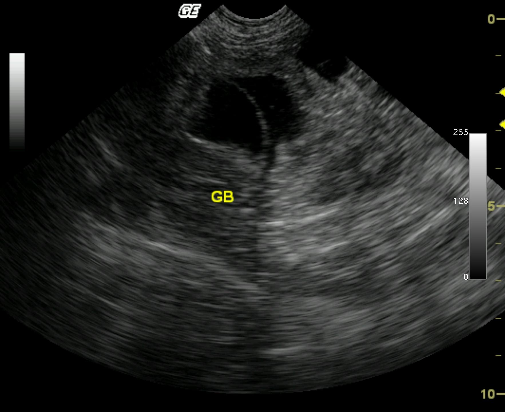
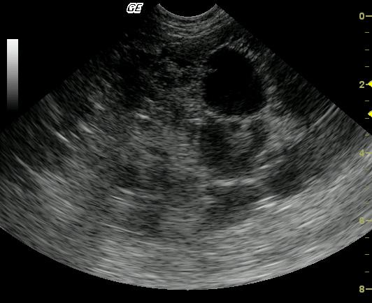
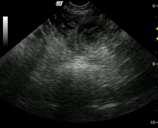
Liver lobe torsion, severe, diffuse, subacute necrohemorrhagic liver infarct in a 8 year old MN DLH cat
History
Comments
No video is available on this patient.
Clinical Differential Diagnosis
Multi-organ pathology: Neoplasia, pancreatitis with sequestrum or abscess, IBD, GI obstruction, hepatic cyst.
DX
Sampling
Abdominocentesis revealed only sterile cystic fluid with blood. Given the intense pain, rapidly declining clinical status, and risk of fatal hemorrhage, rapid surgical intervention was undertaken for left liver lobectomy after survey thoracic radiographs did not reveal any metastatic lesions. Liver lobe torsion was the surgical diagnosis. Histopathologic analysis demonstrated severe, diffuse, subacute necrohemorrhagic infarct with underlying cystadenoma or hepatoma. No malignant tissue was seen by the pathologist on multiple tissue sections.
Sonographic Differential Diagnosis
The hepatic findings are suggestive of a benign or low-grade malignant neoplasm, most likely hepatocellular carcinoma. However, peritoneal effusion is an uncommon accompanying finding, and a secondary or underlying disease process is strongly suspected. Liver lobe torsion possible
Image Interpretation
A pedunculated echogenic, heteroechoic mass is limited to and completely involved the left medial liver lobe. A small to moderate amount of anechoic peritoneal effusion is also present. Free fluid was noted; given the anemia then hemoabdomen is suspected
UA Specific Gravity Range
Outcome
Three-week follow-up revealed a normal physical state and no abnormality on blood chemistry profile. A 4-month follow-up examination was normal as well.
Patient Information
Blood Chemistry
- ALT (SGPT), High
- AST (SGOT), High
- Azotemia
- Creatinine, High
CBC
- RBC, Low
Clinical Signs
- Diarrhea
- Lethargy
- Vomiting
Exam Finding
- Palpable mass
Urinalysi
- Blood Present
- Specific Gravity High
Images