Neoplasia of epithelial origin diagnosed by abdominal fluid analysis in a 10 year old FS mixed breed dog.

History

A 10-year-old FS mixed breed dog was examined for anorexia and lethargy. Weight loss and muscle atrophy were noted on physical exam. Preoperative blood work revealed azotemia, hypoalbuminemia, and hyperglobulinemia. CBC found a low hematocrit, low RBCs, and a neutrophilia. PT and PTT were both within normal reference range.

A 10-year-old FS mixed breed dog was examined for anorexia and lethargy. Weight loss and muscle atrophy were noted on physical exam. Preoperative blood work revealed azotemia, hypoalbuminemia, and hyperglobulinemia. CBC found a low hematocrit, low RBCs, and a neutrophilia. PT and PTT were both within normal reference range.

Comments


Working through ascites by the sonogram is very simple if you just reason through it a bit and ask, “What does this?” If the albumin is low, but greater than 1.5, look for other contributing causes (portal hypertension, lymphatic obstruction/strangulation, imflammatory fluid, neoplasia) to help third space the fluid. If the albumin is less than 1.5 g/dl, third spacing of fluid (trans/mod transudate) can occur spontaneously due to poor oncotic pressure. In this case, the cause of ascites is either protein-losing nephropathy (check urinalysis for significant proteinuria, for example 1.015 USGH and 3+ proteinuria), diffuse disease in the liver causing poor albumin production, or protein-losing enteropathy and/or lymphangectasia (look for mucosal striations). If there are no bleeding masses (spleen hsa or similar) and no ruptured organs (i.e. GI, pancreatic or prostatic abscess), then check the liver for diffuse disease short of overt liver failure (portal hypertension and ascites) and hepatic vein and caudal vena cava dilation. Often I check the liver first when there is ascites (especially if I know its transudate or mod transudate). If the hepatic vein and caudal vena cava are dilated, then the problem is cranial in the thorax (pericardial effusion or right atrial mass, caudal mediastinal neoplasia obstructing the caudal vena cava, or thoracic caval thrombosis). If all of these pathologies described are ruled out in the sonogram then usually the ascites is from neoplasia (carcinomatosis/lymphomatosis), such as in this case with Lucky the dog (classic pancreatic carcinomatosis in dogs or more often elderly cats). The carcinomatosis/lymphomatosis presentation often manifests as a bundle of badness composed of nodular omentum coalesced like cotton candy around a hypoechoic pancreas or lymph nodes. There are often only minor pancreatic changes, however nothing will be naturally curvilinear as in a normal organ. Search PLN, PLE, cirrhosis, carcinomatosis or lymphomatosis in the SonoPath basic search for visual examples of these diseases. Bottom line don’t name your dog Lucky…its never a good thing.


Clinical Differential Diagnosis

Azotemia – prerenal, renal. Hypoalbuminemia – protein losing nephropathy (glomerulonephritis, amyloidosis) or enteropathy (inflammatory bowel disease, neoplasia such as lymphoma, adenocarcinoma, leiomyoma, leiomyosarcoma or mast cell tumor).

DX

Malignant neoplasm of epithelial origin (adenocarcinoma, carcinoma)

Sampling

Cytology results of the fluid aspirated with abdominocentesis and immediate spin down and slide prep of the sediment revealed a malignant neoplasm of epithelial origin (adenocarcinoma, carcinoma).

Sonographic Differential Diagnosis

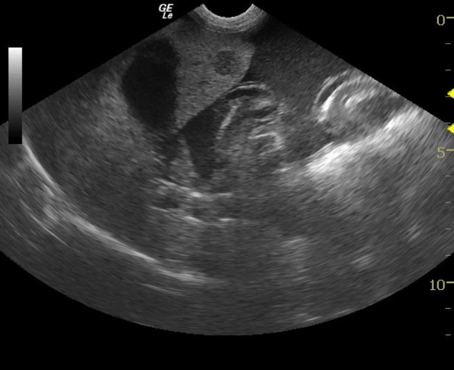
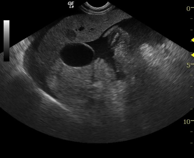
The nodular change in the liver is suggestive of nodular hyperplasia or primary (hepatocellular and choangiocellular) or secondary neoplasia (pancreatic, intestinal, bone marrow, lymphosarcoma and hemangiosarcoma.) The enlarged lymph nodes have a lacy appearance supportive of lymphosarcoma, but cytology would be required. Reactive nodes are a differential, but are less likely than infiltrative neoplasia.

Image Interpretation

The liver was diffusely hyperechoic and coarse, mildly irregular capsule in some areas, with multifocal discrete hypoechoic nodular change. There was a severe accumulation echogenic effusion causing displacement of the liver lobe away from the diaphragm and a well-defined diaphragmatic lung interface. The sublumbar lymph nodes were enlarged, hyperechoic and lacy.

Outcome

The patient was humanely euthanized due to poor prognosis.

Video

Patient Information

Patient Name : Lucky D
Age : 10 Years
Gender : Female, Spayed
Species : Canine
Liz Wuz Here : Yes
Status : Complete
Code : 11_00009

Blood Chemistry

  • Albumin, Low
  • Azotemia
  • Globulin, High

CBC

  • Hematocrit, Low
  • Neutrophils, High
  • RBC, Low

Clinical Signs

  • Anorexia
  • Lethargy

Exam Finding

  • Muscle Wasting
  • Weight loss

Images

liver_nodule_10072010092344liver_nodule_2

Skip to content