A 5-year-old male neutered mixed breed dog was presented for evaluation of sudden onset collapse, hypothermia, and marked dyspnea.
A 5-year-old male neutered mixed breed dog was presented for evaluation of sudden onset collapse, hypothermia, and marked dyspnea.
Right ventricular hypertrophy and pulmonary hypertension due to pulmonic stenosis in a 5 year old MN mixed breed dog
History
Clinical Differential Diagnosis
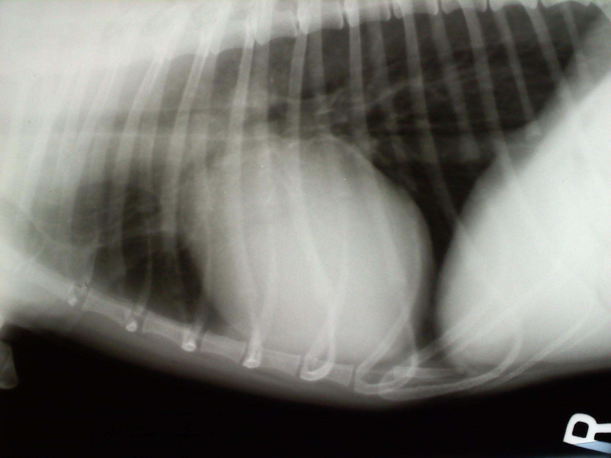
Cardiac – cardiac tamponade, ruptured chordae tendina, cardiomyopathy (dilated/hypertrophic), aortic outflow obstruction. Pleural effusion. Respiratory – pulmonary embolus, lung lobe torsion, edema. Red cells – acute severe anemia, carbon monoxide toxicity.
DX
Sampling
None
Sonographic Differential Diagnosis
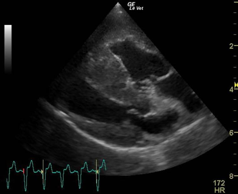
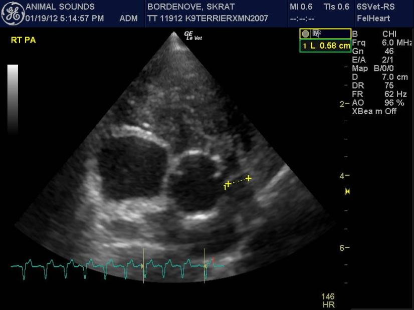
The echocardiogram shows marked to severe right concentric ventricular hypertrophy with a small left ventricular chamber, mild right atrial enlargement, consistent with a pressure overload to the right ventricle alone. Pulmonary hypertension most often results in both a pressure and volume overload to the right heart. The appearance of the right ventricle is likely due to pulmonic stenosis with the elevations in RVOT pressures. This is the likely source of the episode noted, due to reduced cardiac output. The condition appears well advanced with the changes seen here but the heart appears to be compensating at this time with the only mild changes to the right atrial size.
Image Interpretation
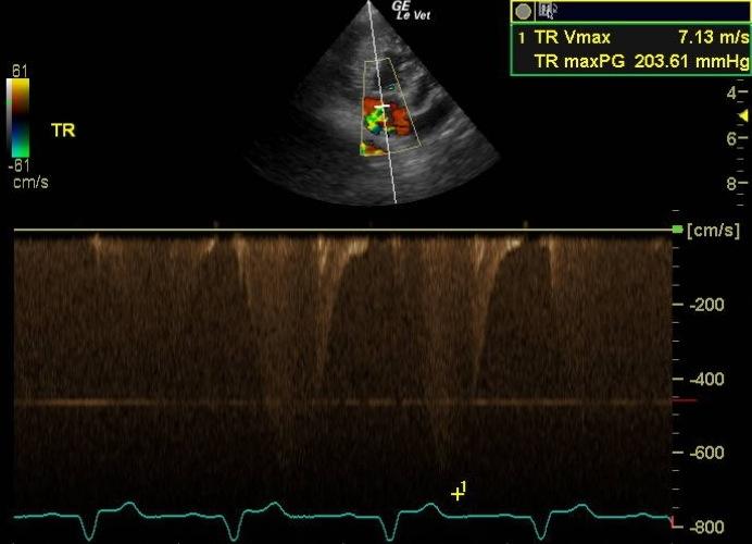
All valves viewed appear normal although the pulmonary artery is narrowed at the level of the pulmonic valve. The left ventricular cavity is small in diastole (1.25 cm) and systole (0.4 cm) with an elevated fractional shortening (0.68 %). There is flattening of the interventricular septum and paradoxical septal wall motion on M-mode exam, consistent with right ventricular hypertension. The left ventricular walls are relatively hypertrophied (0.75 cm septal wall and 0.75 cm free wall). The right ventricle appears markedly to severely hypertrophied and the right atrium appears mildly enlarged in size. The right ventricular outflow tract (RVOT) velocity is markedly to severely elevated at about 6 m/sec, describing a pressure gradient of 144 mmHg. There is mild tricuspid regurgitation, also with a markedly to severely elevated velocity of 7.13 m/sec, describing a pressure gradient of over 200 mmHg. There is also mild pulmonic insufficiency on Doppler exam.
Outcome
The patient expired three months after the echocardiogram in acute respiratory distress.


