10_00073 Fletch T Cranial mediastinal mass, abdominal lymphadenopathy

History

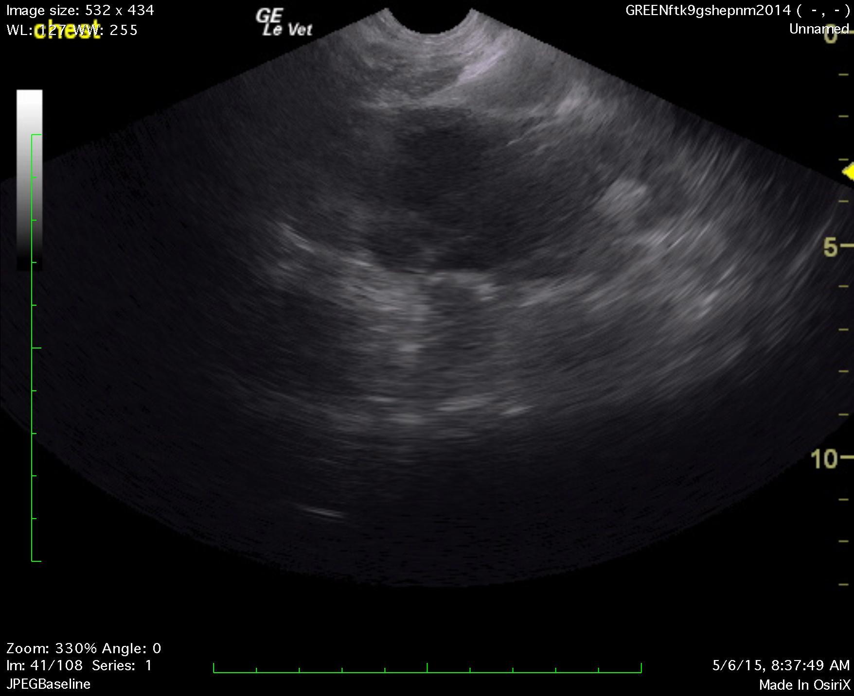
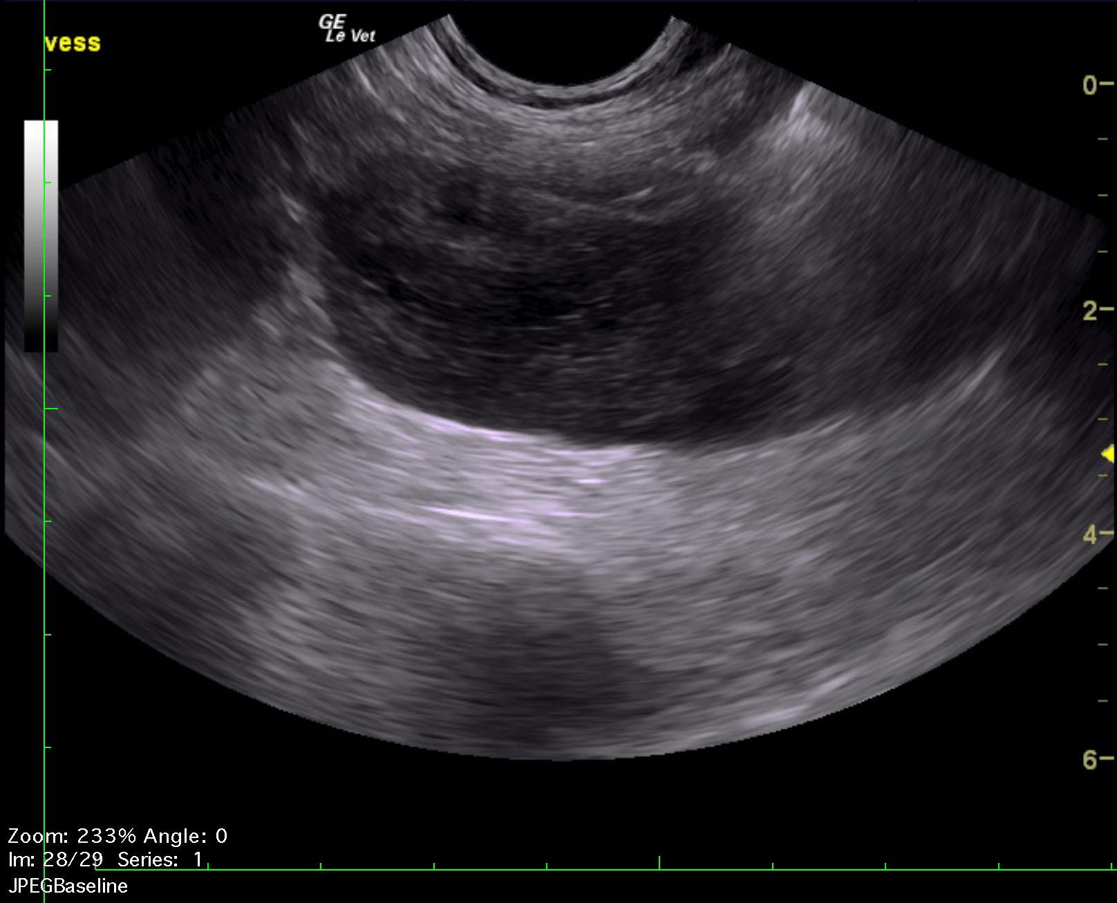
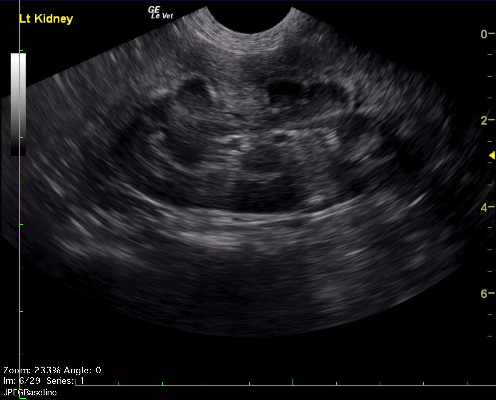
A 1-year-old NM German shepherd with a history of PU/PD and hypercalcemia was presented for evaluation of lethargy, weight loss, and anorexia. Left-sided nephromegaly was present on survey radiographs. Abnormalities on serum biochemistry were hypercalcemia, azotemia, and mildly elevated ALT activity.


Clinical Differential Diagnosis

Hypercalcemia of malignancy – lymphoma, carcinoma
Fungal disease
Kidney disease secondary to the hypercalcemia

DX

Multicentric LSA, suspect

Sampling

None

Sonographic Differential Diagnosis

Multicentric lymphadenopathy involving cranial mediastinum and mid caudal abdomen.
Medullary rim kidney, potentially owing to hypercalcemia or emerging neoplasia within the kidney.
FNA of the lymph node masses and cranial mediastinal mass is recommended. Hypercalcemia is likely owing to malignancy. Lymphoma or similar neoplasia is suspected.

Image Interpretation

The chest in this patient presented a cranial mediastinal mass. This was comprised of multiple lymph nodes. A mid-abdominal lymph node mass was noted and measured 4 x 3 cm. There are separate, mixed hypoechoic mid-abdominal lymph node masses. The kidneys were swollen in this patient. The left kidney measured 7.4 cm with medullary rim sign and loss of corticomedullary detail. The right kidney revealed similar changes to the left kidney. The right kidney measured 7.3 cm. Pyelectasia of both kidney was noted.

Outcome

None

Video

Patient Information

Age : 1 Year
Gender : Male, Neutered
Species : Canine
Status : Complete

Blood Chemistry

  • ALT (SGPT), High
  • Azotemia
  • Calcium, High

Clinical Signs

  • Anorexia
  • Lethargy
  • PU-PD
  • Weight loss

Exam Finding

  • Large Kidneys

Images

_20150627104703fletch_talbot_hypercaclemia_lsa_ln_2fletch_talbot_hypercaclemia_lsa_lk_2

Skip to content