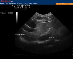
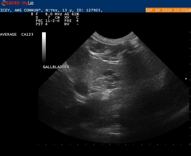
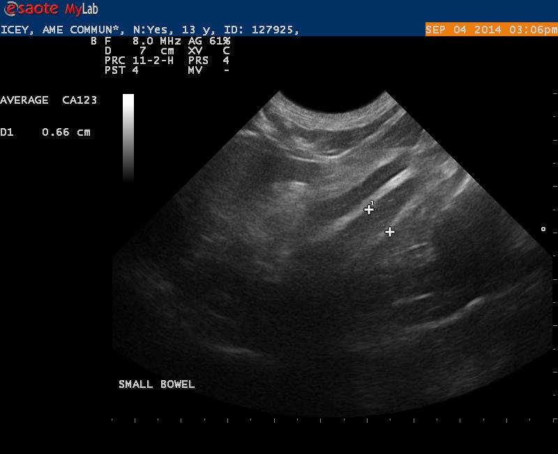
Icey is a 13 year old Maine Coon with a recent history weight loss, vomiting and loss of appetite.
I have 3 questions:
1. Is this a double walled gallbladder?
2. What is the source of the hypoechoic branching. This area was not vascular per color doppler
3. Is the image labeled “small bowel” actually bowel that is thickened with loss of normal layering?
Sorry to be posting so much, but I am starting to get more active and I am seeing pathology now that I can’t always interpret.
12 responses to “Dual Galbladder”
You actually have a dual gb
You actually have a dual gb … the sonographers 6 toed cat.. genetic anomaly not pathological. The parenchymal cystic lesion is a biliary adenoma benign tumor in oplder cats just watch if gets locally expansive. Neither are clinical players but here is a series of double gb and double layered gb…..
The first is from the atlas 400 pathology CD case 57 where this dual GB cat also formed a Gb mucocele and mild cholecystitis in the vestigial gb or vice versa…
Or a double Gb cat with
Or a double Gb cat with cholecystitis
Or a double Gb cat with
Or a double Gb cat with cholecystitis
Non inflammatory double
Non inflammatory double layered GB: Here is a dog with a true double layered GB from ascites formation… not inflammatory.
Inflammatory double layered GB: Then a chronic cholecystitis double layered wall of a dog with GB mucocele and chronic cholecystitis.
Then a normal cat with a normal dual GB but not double layered…
Do you have a video of
Do you have a video of the “bowel”?? I can’t say on the still if this is bowel or adjacent pv or duct….
Do you have a video of
Do you have a video of the “bowel”?? I can’t say on the still if this is bowel or adjacent pv or duct….
Thanks EL. I don’t have a
Thanks EL. I don’t have a cine- but I will try to get the cat back in. I suppose I should have followed the segment till I could show normal bowel.
Thanks EL. I don’t have a
Thanks EL. I don’t have a cine- but I will try to get the cat back in. I suppose I should have followed the segment till I could show normal bowel.
yes in general transitions
yes in general transitions form abnormal to normal and vice versa have the most diagnostic information.
take a look at this to help further rgearding the scanning, recognition, and interpretation in a laid back place:
http://sonopath.com/forum/sonopath-scanning-clinical-pathology-seminar-feb-19-22-2015-puerto-rico-drs-vet-techs