RADS – Hypovolemia and Underperfusion of the Lung with Atalectasis in a 16 year old FS Siamese cat

History

History: 16-year-old F/S Siamese being boarded but on presentation owner said that she has been eating alot, vomiting about 10 days, had some diarrhea;  lost “alot” of  weight in the last couple of weeks. 

Physical Exam: thin, not grooming herself, lungs clear, no peripheal lymph enlargement, eyes sunken in but clear

CBC: HCT decr 25.3%; Hgb decr 8.7; reticulocytes 64.4; WBC 19.54; neutrophils 15.07; EOs incr 1.99

Chemistry: electrolytes – sodium incr171 and potassium incr 5.9;  BUN incr 69, CREA incr 2.9 (dehydration?) ; T4 0.8 is a low-end of normal

Comments

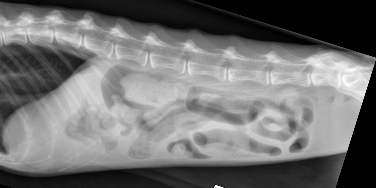
Although there is some overlap with neoplastic infiltrate in general this is very unlikely in this case. The lungs further presented age related changes and plaque like atelectasis. The radiographs do not identify any evidence of obstructive intestinal pathology but are suggestive of a funnctional intestinal ileus. Abdominal ultrasound is recommended to further assess the intestinal tract and remainder of the abdominal organs.

DX

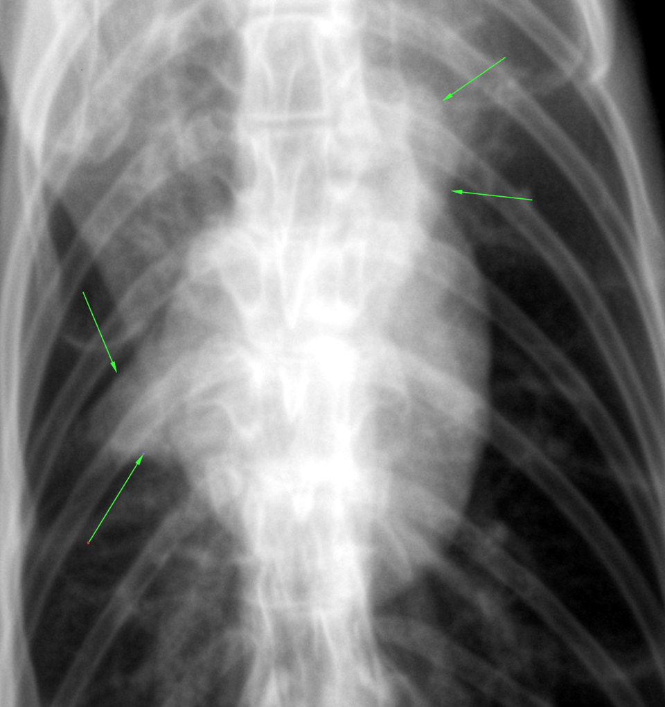
hypovolemia and underperfusion of the lung. There was residual fat versus focal atelectasis in the area of the right middle lobe.

Image Interpretation

right lateral, left lateral and VD thorax and abdomen – Osseous structures: There were moderate degenerative changes
associated with the axial sceleton.
Extrathoracic/-abdominal soft tissues: Within normal limits.
Abdominal Structures:
The serosal detail was severely reduced.
The liver, spleen, kidneys and urinary bladder were not accessible. The stomach contained a mild amount of gas. The small intesinal loops
were mildly dilated with gas but of even diameter. The large intestine
contained a moderate amount of fecal material and gas. There was no
obstructive pattern noted.
Intrathoracic structures:
The upper cervical esophagus contained a mild amount of gas which is a
normal incidental finding.
The course of the trachea was normal.
There was no mediastinal widening and no evidence of mediastinal
lymph node enlargement. There was no mediastinal shift.
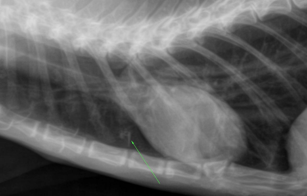
The cardiac silhouette was small. The major and pulmonary vessels were
thin. There was redundancy of the aortic arch which is a normal
incidental finding.
The lungs presented a hypovascular pattern and a mild generalized
bronchial pattern which was likely age related. There was a sharply
delineated triangular shaped soft tissue opacity silhouetting with the right
cardiac border in the ventrodorsal view.
A geometrical shaped well delineated soft tissue opacity was seen cranial
to the cardiac silhouette.

Patient Information

Patient Name : Aichan Devito, Critical Vet Care
Age : 16 Years
Gender : Female, Spayed
Species : Feline
Liz Wuz Here : Yes
Status : Complete

Blood Chemistry

  • BUN high
  • Creatinine, High
  • Potassium, High
  • Sodium, High

CBC

  • Eosinophils, High
  • Hematocrit, Low
  • Neutrophils, High
  • WBC, High

Clinical Signs

  • Diarrhea
  • Polyphagia
  • Vomiting
  • Weight loss

Exam Finding

  • Poor or unkempt coat
  • Weight loss

Images

bildschirmfoto_2015-10-15_um_13bildschirmfoto_2015-10-15_um_13bildschirmfoto_2015-10-15_um_13

Skip to content