MRI – Cerebullar abiotrophy and mild generalized brain atrophy
History
Comments
T1-weighted pre- and post-contrast, T2-weighted, and FLAIR series are available for review.
DX
Image Interpretation
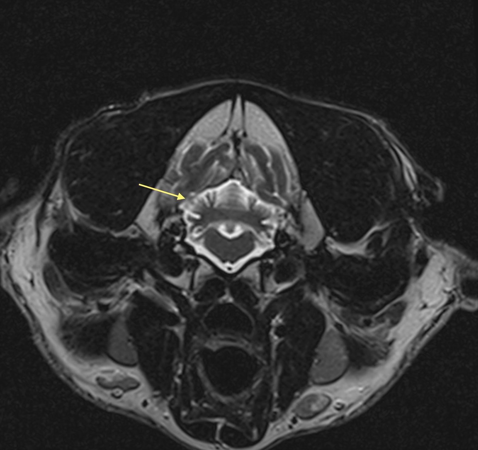
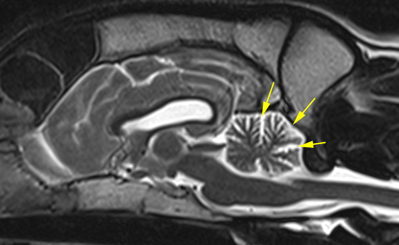
MRI of the head – The development of both the cranial and caudal cranial fossa is within normal limits. The cerebellar parenchyma is decreased in volume in relation to the size of the caudal fossa. The cerebellum is surrounded by a broad rim of cerebrospinal fluid (CSF). The cerebellar foliae are widened and filled with CSF.
The CSF spaces within the cranial fossa persent mild enlargement to. Mild T2-hyperintensity of the cerebral white matter and mild thinning of the cerebral cortex is noted. The sulci and gyri are deep. There is mild bilateral lateral ventricular enlargement. Partial absence of the septum pellucidum is noted.
No abnormal contrast enhancement is noted. The mandibular lymph nodes are rather prominent (up to 7mm in diameter).
Outcome
Possible etiologies of the abiotrophy include:
inhertited storage disease such as adult onset neuronal ceroid lipofuscinosis (NCL)
neuroaxonal dystrophy
and cerebellar cortical degeneration.
There is no indication of inflammatory/infectious pathology.
Genetic testing for NCL is available and should be enforced.
But note: The causative mutations have been identified in English setters, border collies, American bulldogs, and miniature longhaired dachshunds. The commercially available tests are likely not validated for Pit Bulls.
Consider examination of the litter mates!
Pathologic identification of the stored ceroid- or lipofuscin-like lipopigments in affected tissues may be attempted by fundus examination of the eye.
Another diagnostic option would be a brain biopsy.
Video
Patient Information
Images