This 9 year old FS Portugese Water Dog presented with lameness on right front. Painful at the right carpus. History of DJD.
This 9 year old FS Portugese Water Dog presented with lameness on right front. Painful at the right carpus. History of DJD.
This 9 year old FS Portugese Water Dog presented with lameness on right front. Painful at the right carpus. History of DJD.
This 9 year old FS Portugese Water Dog presented with lameness on right front. Painful at the right carpus. History of DJD.
CT of the front limbs, plain – The computed tomography reveals bilateral elbow dysplasia because of a fragmented
coronoid process with advanced degenerative joint disease.
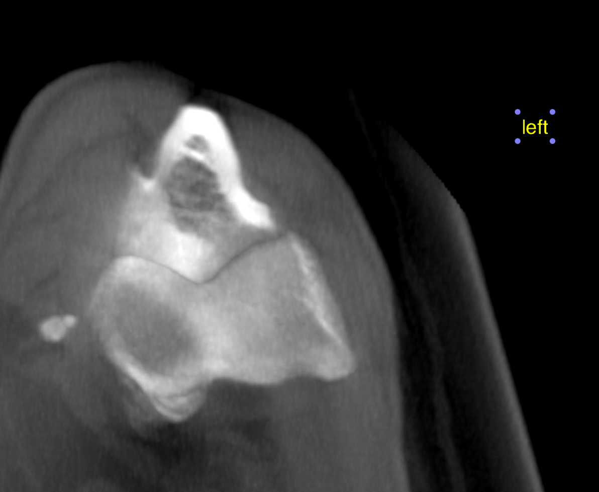
The right medial coronoid process shows a large triangular fragment which is located
in situ at the tip of the process and several small isolated bony fragments which are
mildly displaced into the medial joint compartment. Severe osteophytes are seen at the
periarticular margins. The humeral trochlea presents focal sclerosis and mild flattening
of the subchondral bone. Only minor radioulnar step with long ulna is noted.
The tip of the left medial coronoid process shows marked deformity and heterogenous
opacity. One small and two medium sized fragments are displaced into the medial joint
compartment. Severe osteophytes are seen at the periarticular margins. The humeral
trochlea presents focal sclerosis and mild flattening of the subchondral bone. Only
minor radioulnar step with long ulna is noted.
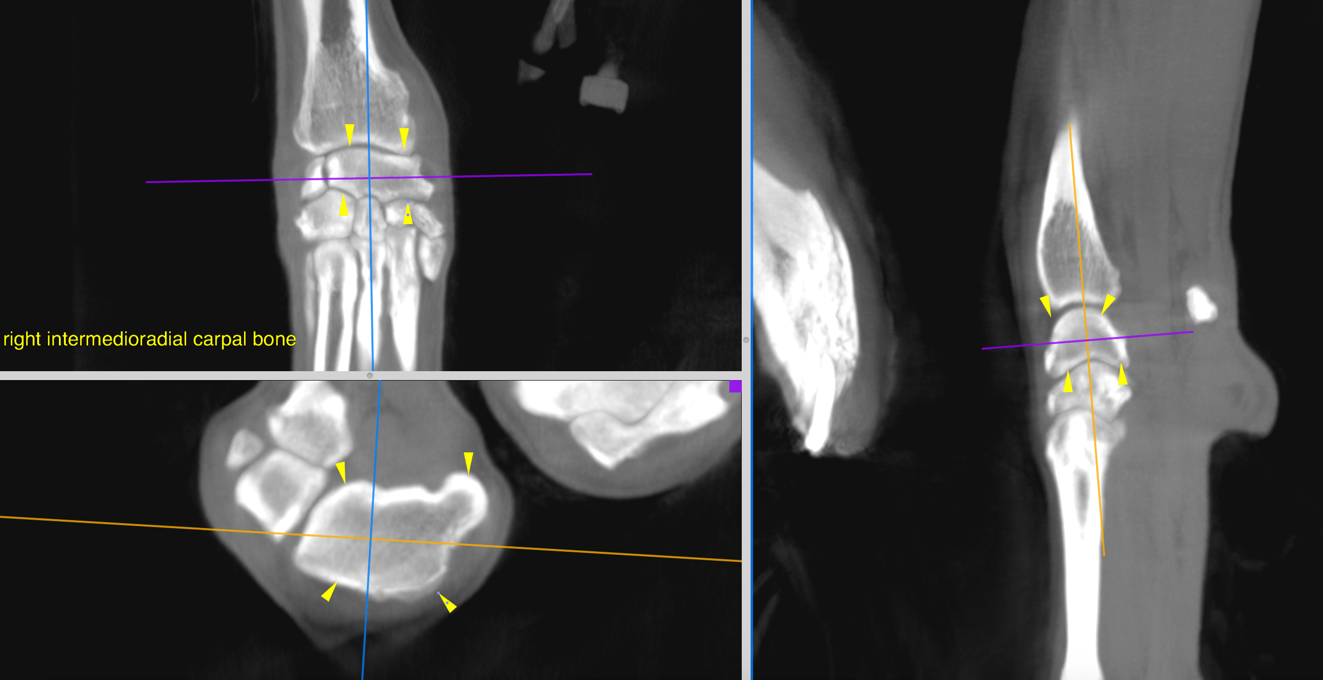
Moderate osteophyte formations are noted at the periarticular margins of the right and
left antebrachiocarpal, intercarpal and carpometacarpal joints. Both medial styloid
processes of the radius present moderate enthesiophytes circumferential to the abductor pollicis longus muscle. The tendon sheath and tendon of the abductor pollicis longus
muscles is within normal limits bilaterally.
There is no abnormal soft tissue swelling and no evidence of a fracture or neoplasia in
the right carpal region.
Both third metacarpophalangeal joints reveal marked chronic osteoarthritis with a
vacuum phenomenon of the right joint which is due to recent joint distraction and an
incidental finding.
There is no evidence of a recent traumatic injury.