This 15 year old intact F Chihuahua dog presented with a 3 day history of vomiting and diarrhea, rDVM concerned about possible collapsed trachea, no history of heart murmur, however heard a heart murmur today, abdomen distended, in respiratory distress
This 15 year old intact F Chihuahua dog presented with a 3 day history of vomiting and diarrhea, rDVM concerned about possible collapsed trachea, no history of heart murmur, however heard a heart murmur today, abdomen distended, in respiratory distress
The radiographic findings are compatible with left-sided congestive cardiac failure and cardiogenic pulmonary edema owing to mitral regurgitation.
Rads of the thorax and abdomen: The patient is obese.
Expected age-related degenerative changes are associated with the skeleton.
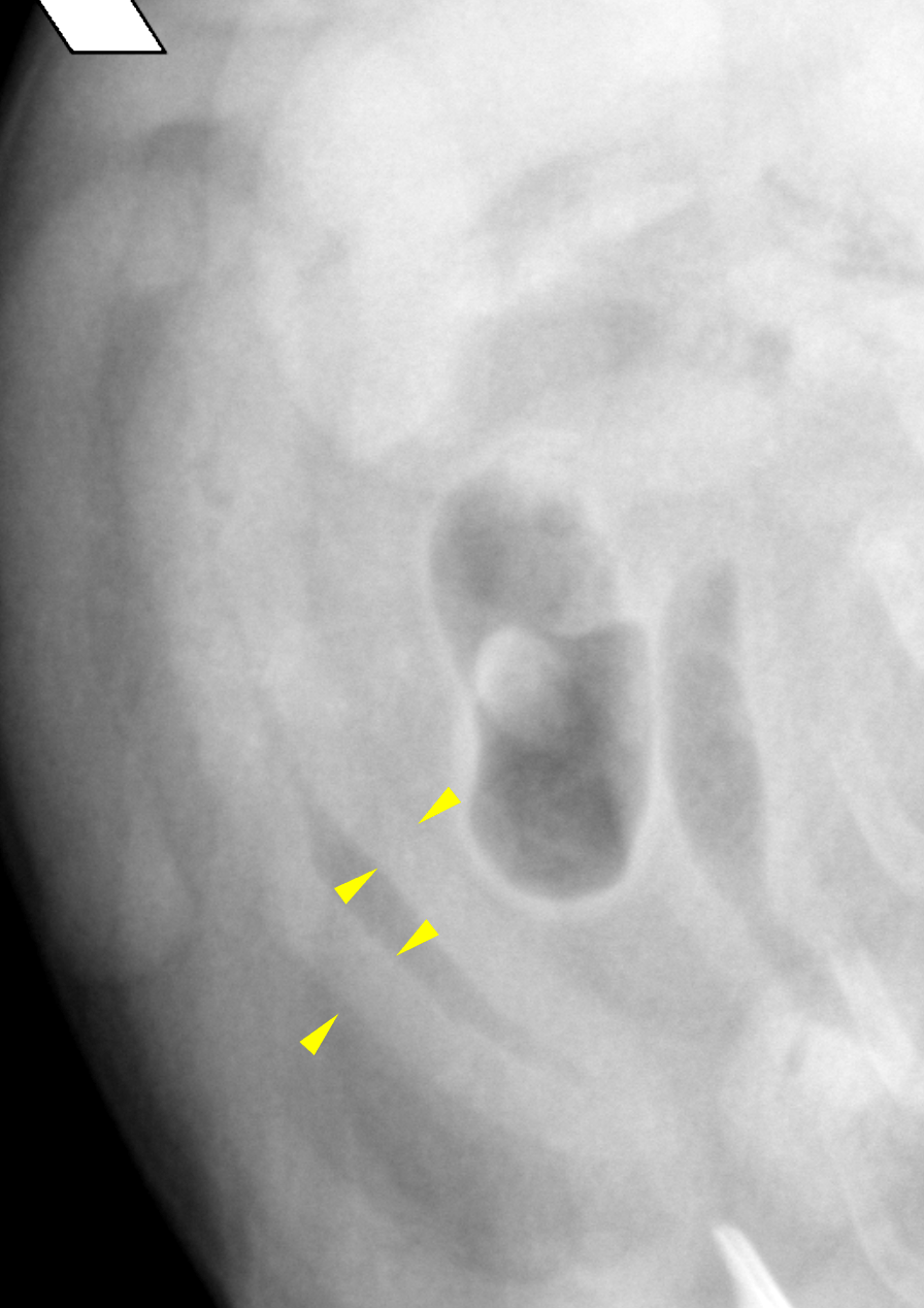
The cardiac silhouette is enlarged in long and short axis presenting a steep caudal
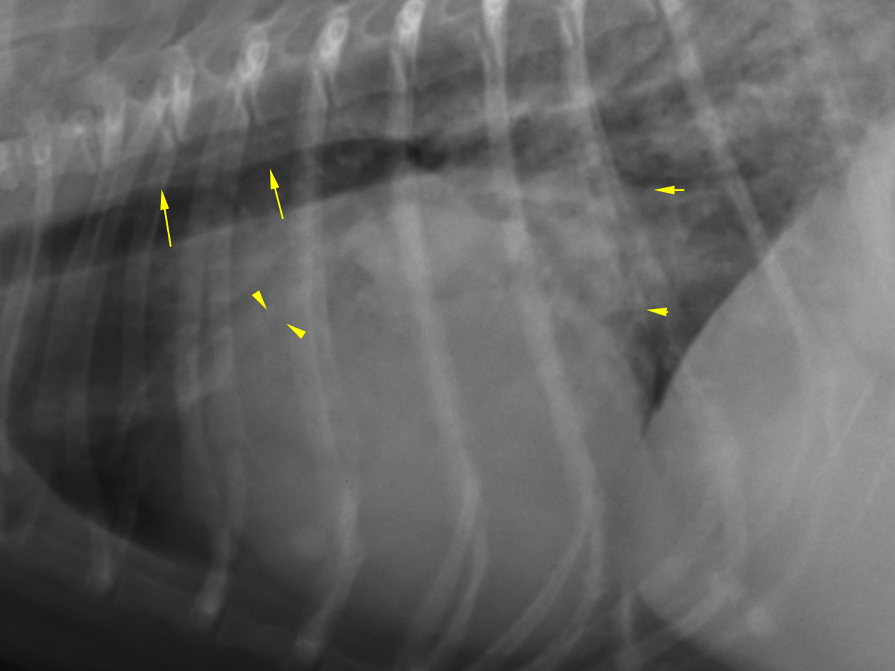
contour with atrial tenting and loss of the caudal cardiac waist. There is splitting of the
mainstem bronchi, and the trachea is elevated. The pulmonary veins are enlarged.
There is a marked perihilar alveolar lung pattern with air bronchograms.
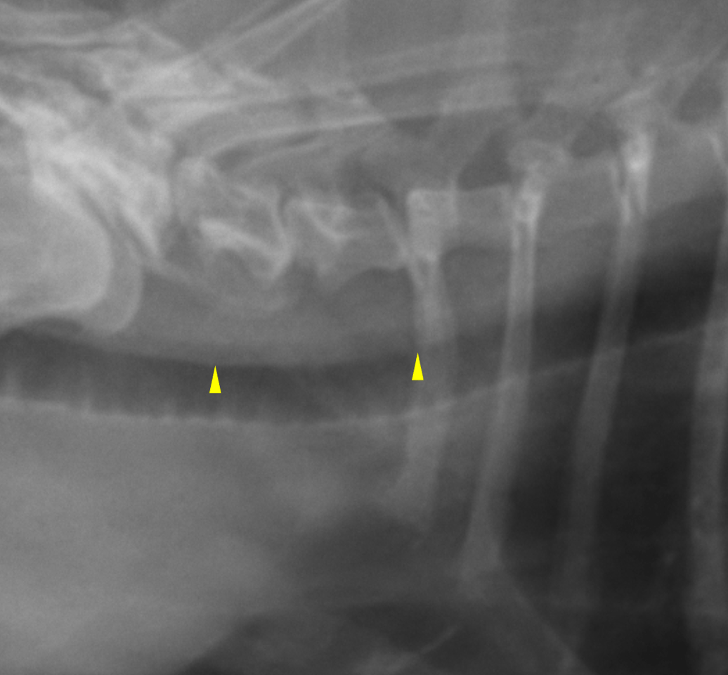
The trachea presents a redundant tracheal membrane, which is an incidental finding, a
true collapse is not noted.
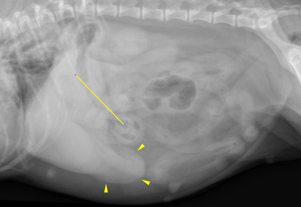
The abdomen is pot bellied.
The liver is moderately enlarged with rounded lobar margins. Small mineral opacities
are associated with the kidneys; otherwise, the kidneys are within normal limits for
size and shape. This gastric axis is rotated caudally as a function of the hepatic
enlargement. The stomach is empty except for a small amount of gas and is
contracted. There is the subjective impression of generalized small intestinal wall
thickening although the assessment of the intestinal wall is limited radiographically in
general.
Overall assessment:
Moderate left-sided cardiomegaly
Pulmonary venous hypertension
Cardiogenic pulmonary edema
Moderate generalized hepatomegaly
Suspicion of small intestinal wall thickening
The most likely
underlying disease is mitral valve endocardiosis/myxomatous degeneration.
The clinically observed coughing is likely owing to the pulmonary edema and
compression of the left mainstem bronchus due to left atrial enlargement. A tracheal
collapse is not apparent on the radiographs.
Consider steroid induced hepatitis, vacuolar hepatopathy, infectious hepatitis or diffuse
infiltrative neoplastic disease as underlying disorder to the moderate generalized
hepatomegaly. In association with the pot bellied appearance of the abdomen
Cushing’s with steroid induced hepatitis is regarded a likely differential diagnosis.
As mentioned above the assessment of small intestinal wall thickness is limited by
means of radiographs, but in correlation with the with the vomiting reported in the
history of primary or secondary small intestinal pathology should be considered and
further workup by means of abdominal ultrasound is warranted.
A full cardiac echo and abdominal ultrasound are recommended for further definition.
Diuretic treatment is indicated after staging of the cardiac disease.