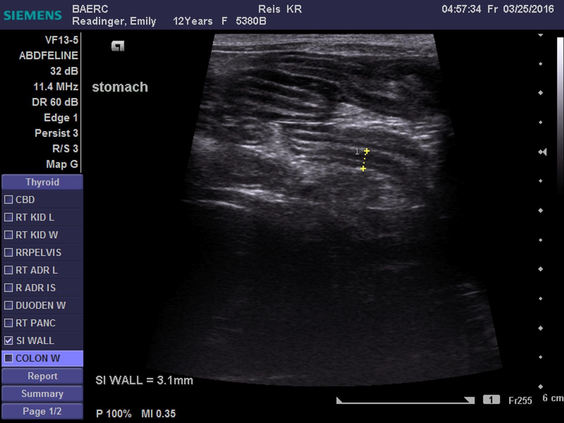
This 12 year old FS DSH cat presented for vomiting, lethargy and anorexia of 4 days duration.
Physical Exam: Depressed, lethargic,weight loss, grade 1-2/6 systolic ht mm, severely prolonged skin turgor, appr 10% dehydrated, tacky mm, cachexic, generalized muscle atrophy, doughy potbellied abdomen, poor coat, thyroid slip palpated and severe dental disease.
CBC: severe leukocytosis(43,000) , neutrophilia(33,000), mild lymphocytosis and monocytosis, HCT-41%
Chemistry: BG=621, BUN-73, creat-1.5, phos-6.4, Ca-8.8, TP-7.1, t. bili-1.1
Urine Analysis: SG=1.028, ph-6, 3+ serum ketones, lg amount of glucose, trace protein
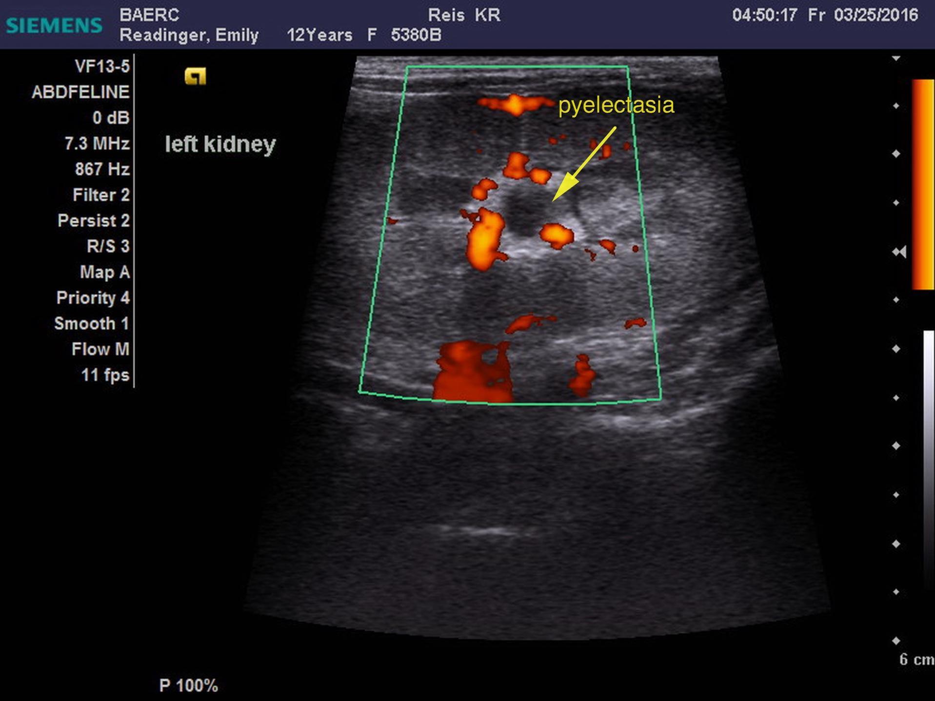
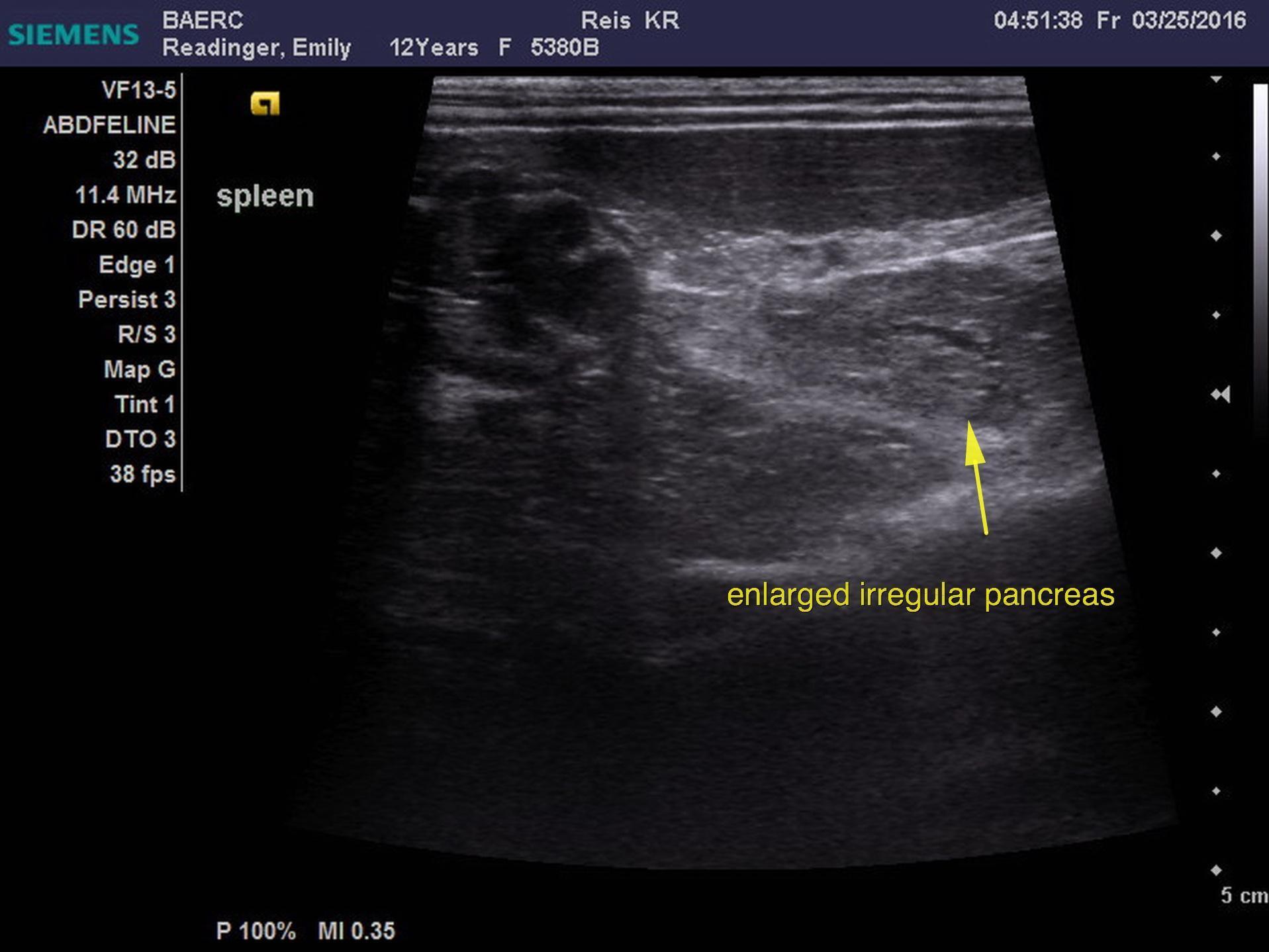
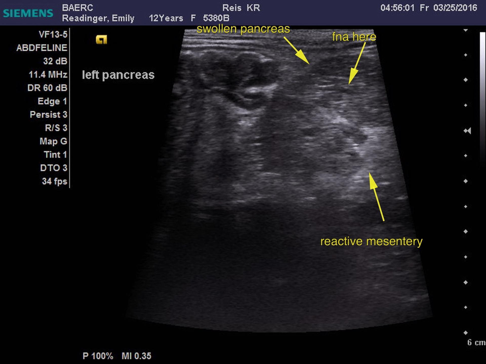
FPLI-abnormal, T4=0.5 low. Blood gas: ICa-1.11 slightly low, BE=12,lactate=0.63, pH=7.47, this blood gas was performed 48hr post admit.