This 2 year old MN Pit Bull Terrier presented with left hind lameness, worse after excercise, of 6 months duration
Physical exam: subtle laxity of the left stifle.
This 2 year old MN Pit Bull Terrier presented with left hind lameness, worse after excercise, of 6 months duration
Physical exam: subtle laxity of the left stifle.
• Chronic degenerative joint disease (DJD) of the left stifle with complete failure of the cranial cruciate ligament and moderate osteoarthritis • Chronic DJD of the right stifle with indirect evidence of cranial cruciate ligament (CCL) failure and mild osteoarthritis • Moderate bilateral hip dysplasia • Asymmetric disuse atrophy of the thigh musculature
Rads-
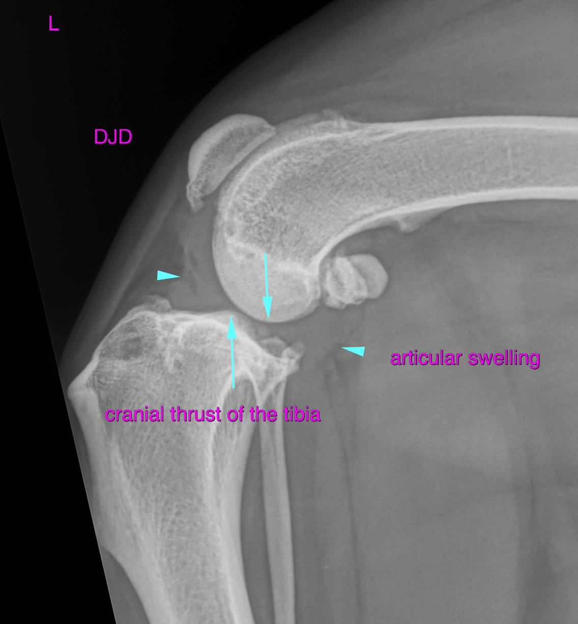
Left stifle :A moderate articular soft tissue swelling is noted. The tibia presents moderate cranial thrust with respect to the mid-point of the femoral condyles. Moderate osteophyte formations are seen at the periartcular margins of the femoropatellar and femorotibial joint.
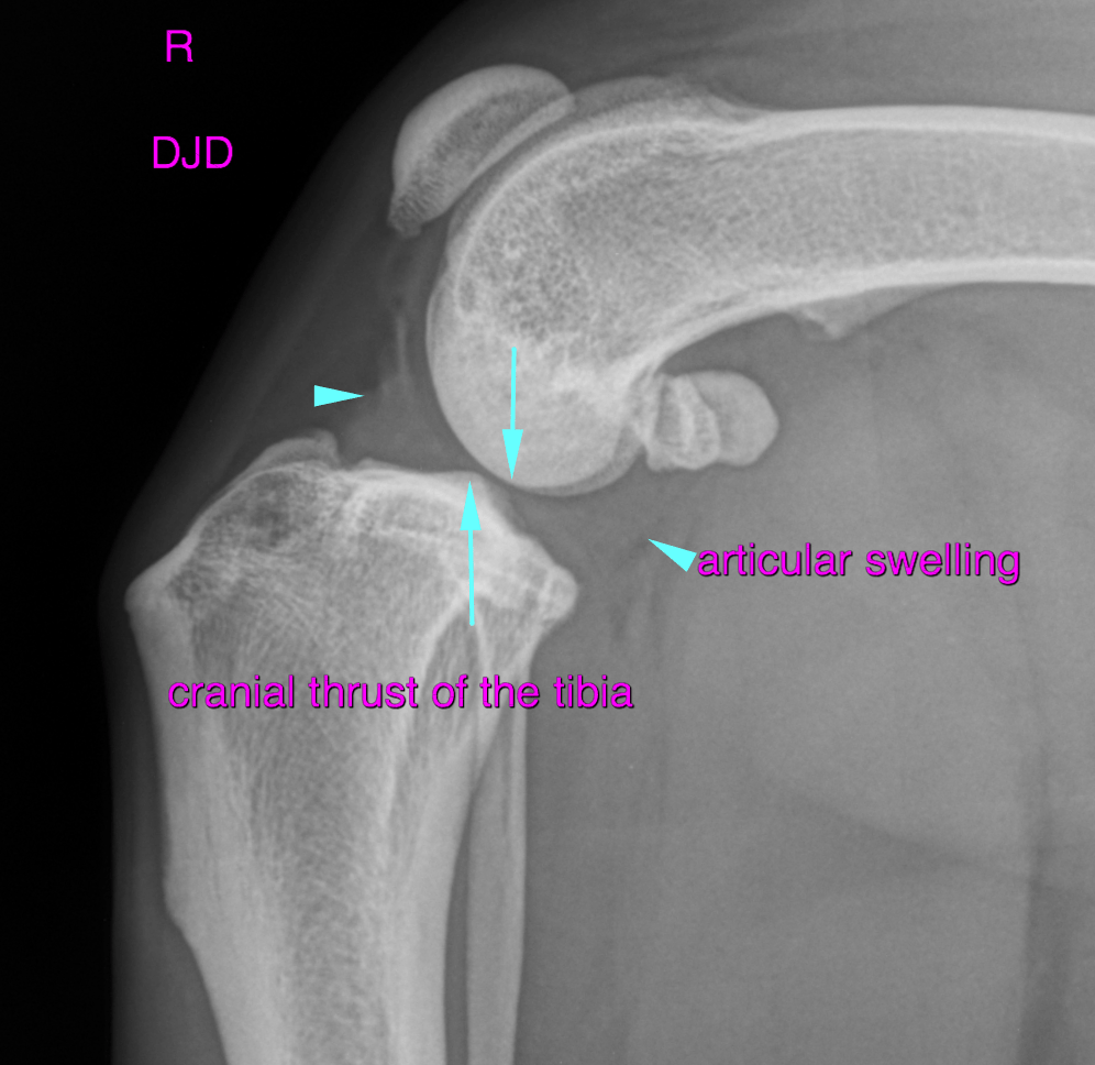
Right stifle: A mild articular soft tissue swelling is noted. The tibia presents mild cranial thrust with respect to the mid-point of the femoral condyles. Mild osteophyte formations are seen at the periartcular margins of the femoropatellar and femorotibial joint.
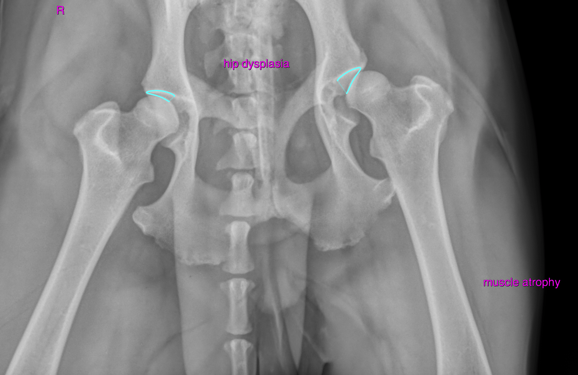
Pelvis: Both hip joints present moderate signs of dysplasia with divergence of the joint spaces and poor coverage of the femoral heads by the acetabular roof. Emerging degenerative joint disease is noted bilaterally. The findings are symmetric between the right and left side. The muscle volume appears to be reduced in both thighs in general. However, the findings are more pronounced for the left rear.
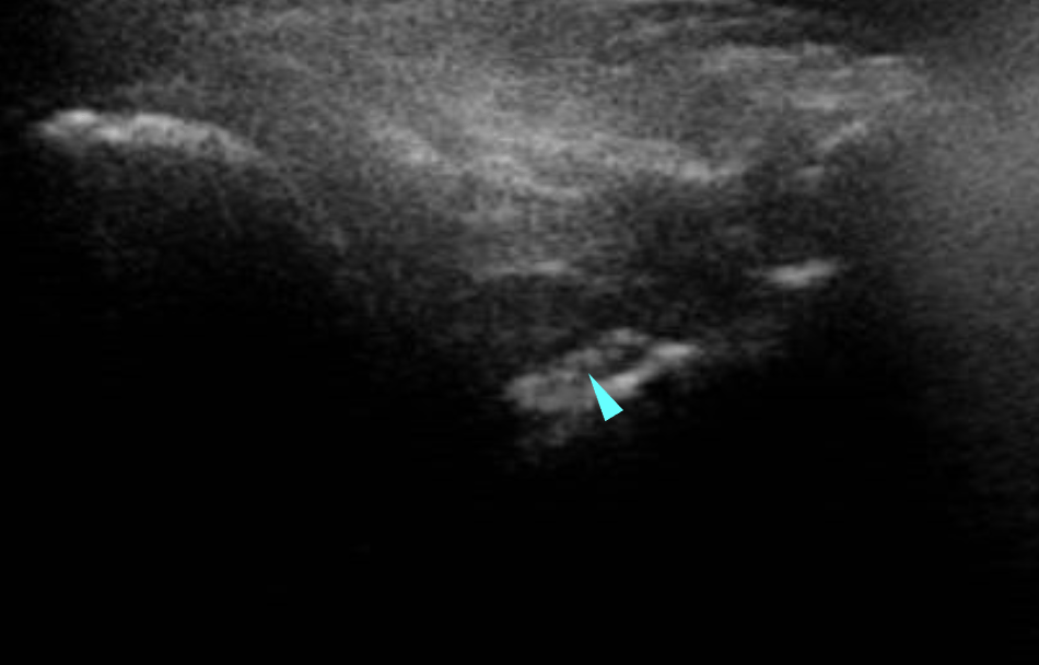
Ultrasound –
Left stifle:
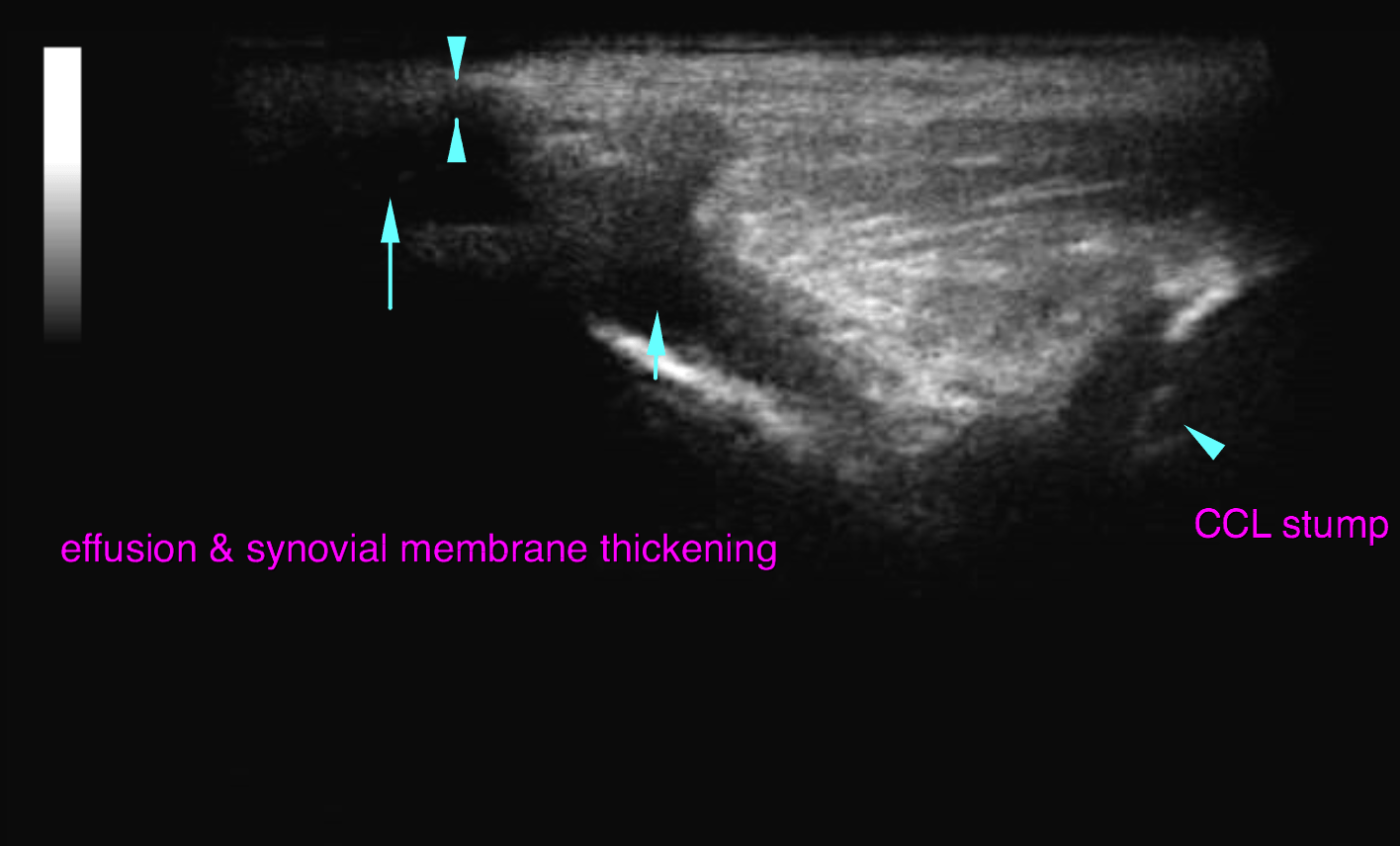
The supra- and infrapatellar recess present a moderate amount of anechoic effusion and synovial membrane thickening respectively. A moderate amount of osteophytes is seen at the periarticular margins. The femoropatellar joint cartilage presents mild irregularity at the most proximal extent of the femoral trochlea. The infrapatellar fat pad presents an irregular echo pattern. The cranial cruciate ligament (CCL) presents as an echogenic stump of fibres at the intercondylar eminence of the tibia surrounded be anechoic periligamentous effusion.