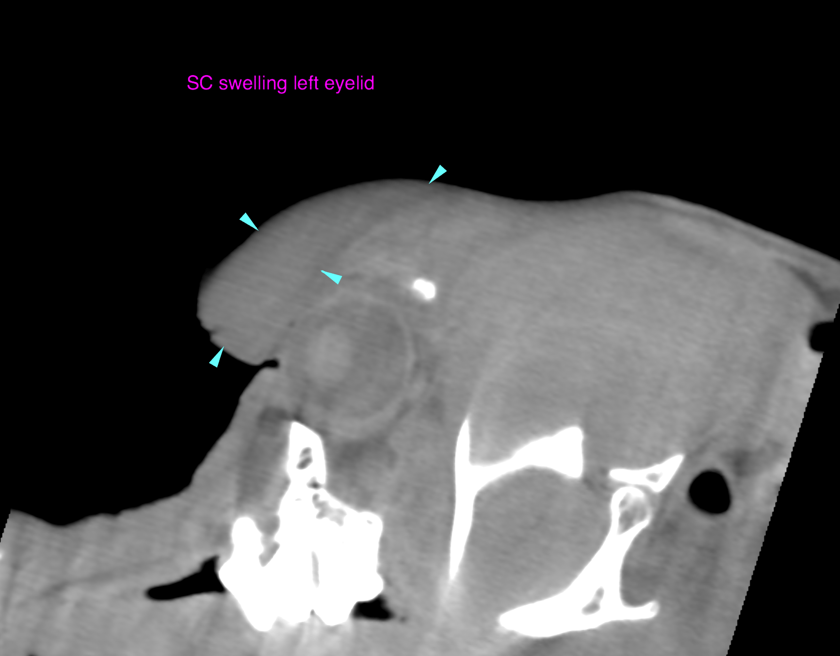
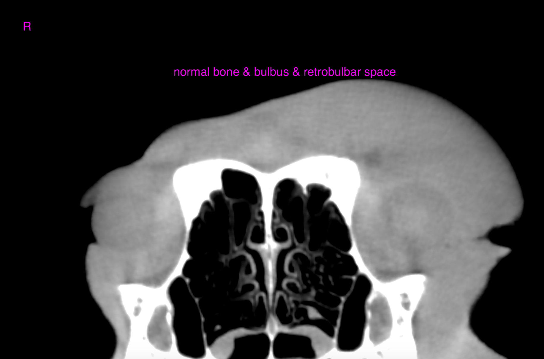
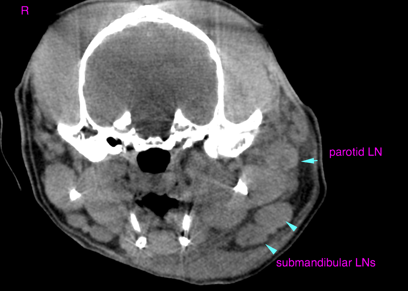
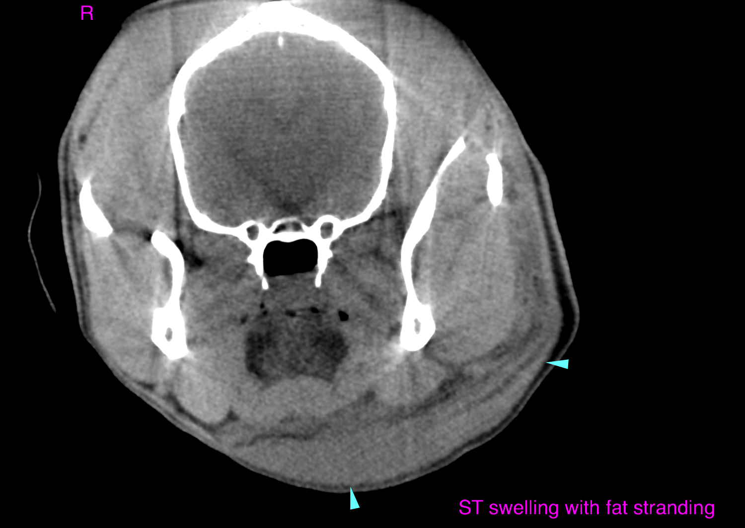
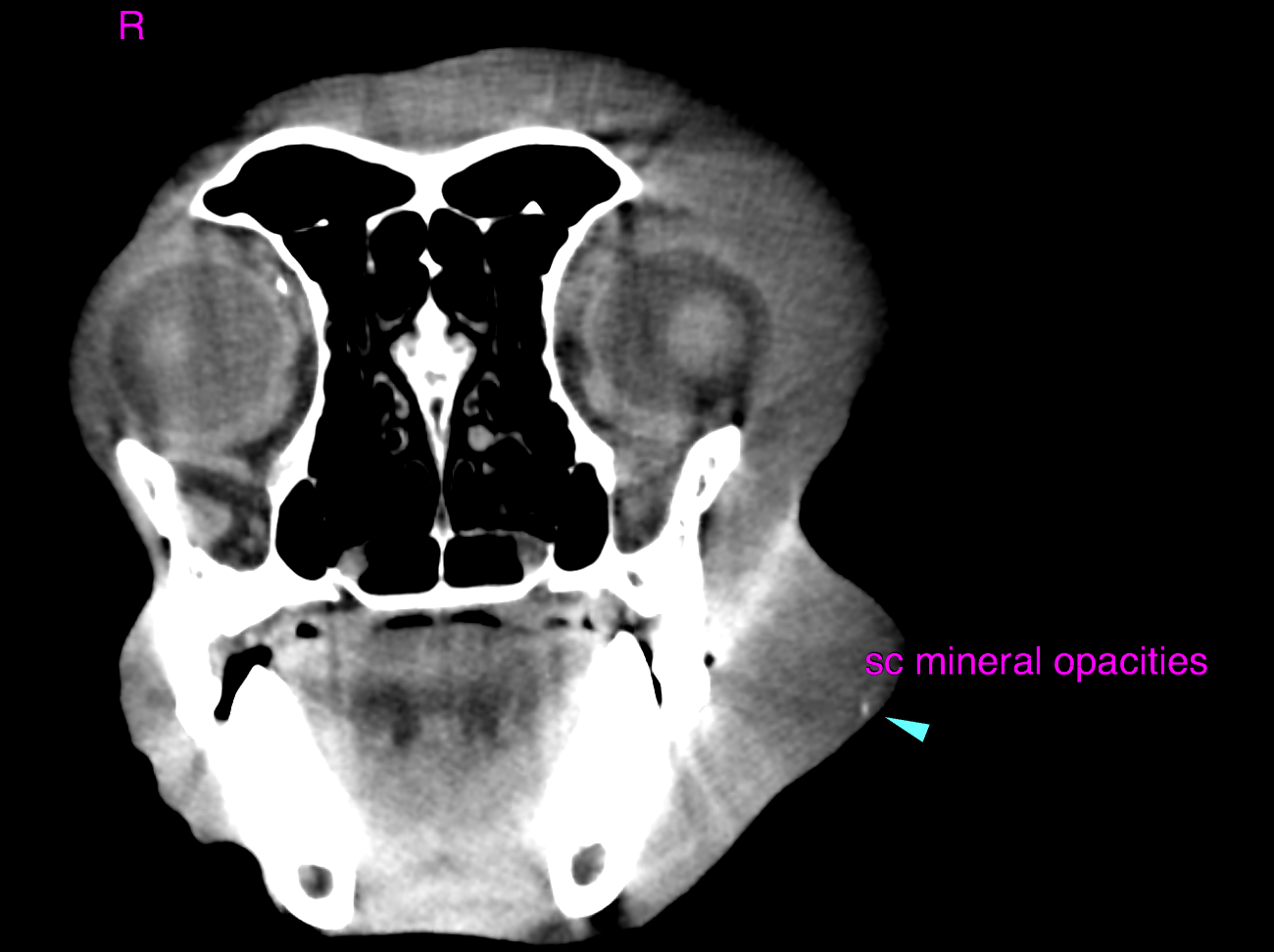
This 7 year old FS Beagle mix presented for progressively worsening swelling around the left eye and down the right side of the face. Cuterebra removed from small puncture on left dorsal eye lid. R/O FB reaction vs cellulitis vs tumor
This 7 year old FS Beagle mix presented for progressively worsening swelling around the left eye and down the right side of the face. Cuterebra removed from small puncture on left dorsal eye lid. R/O FB reaction vs cellulitis vs tumor