Abdominal Mass…..on a Boa Constrictor!

History

SonoPath goes exotic! A 26-year-old Boa Constrictor with an abdominal mass.

Patient managed by Dr. Todd Wolf, Companion AH, Parsippany, NJ, USA and imaged by Dr. Eric Lindquist of SonoPath.com & NJ Mobile Associates, Sparta, NJ, USA.

SonoPath goes exotic! A 26-year-old Boa Constrictor with an abdominal mass.

Patient managed by Dr. Todd Wolf, Companion AH, Parsippany, NJ, USA and imaged by Dr. Eric Lindquist of SonoPath.com & NJ Mobile Associates, Sparta, NJ, USA.

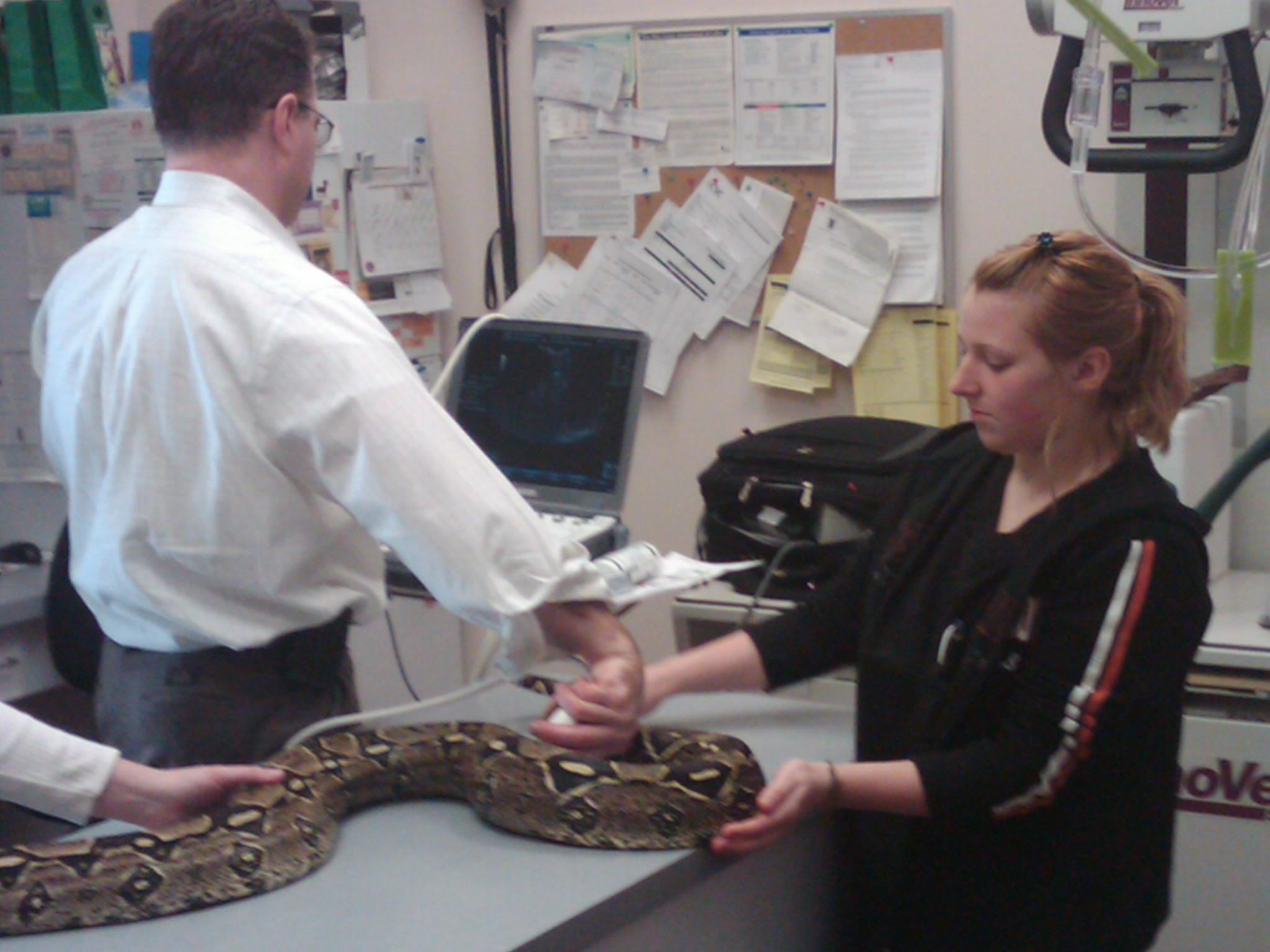
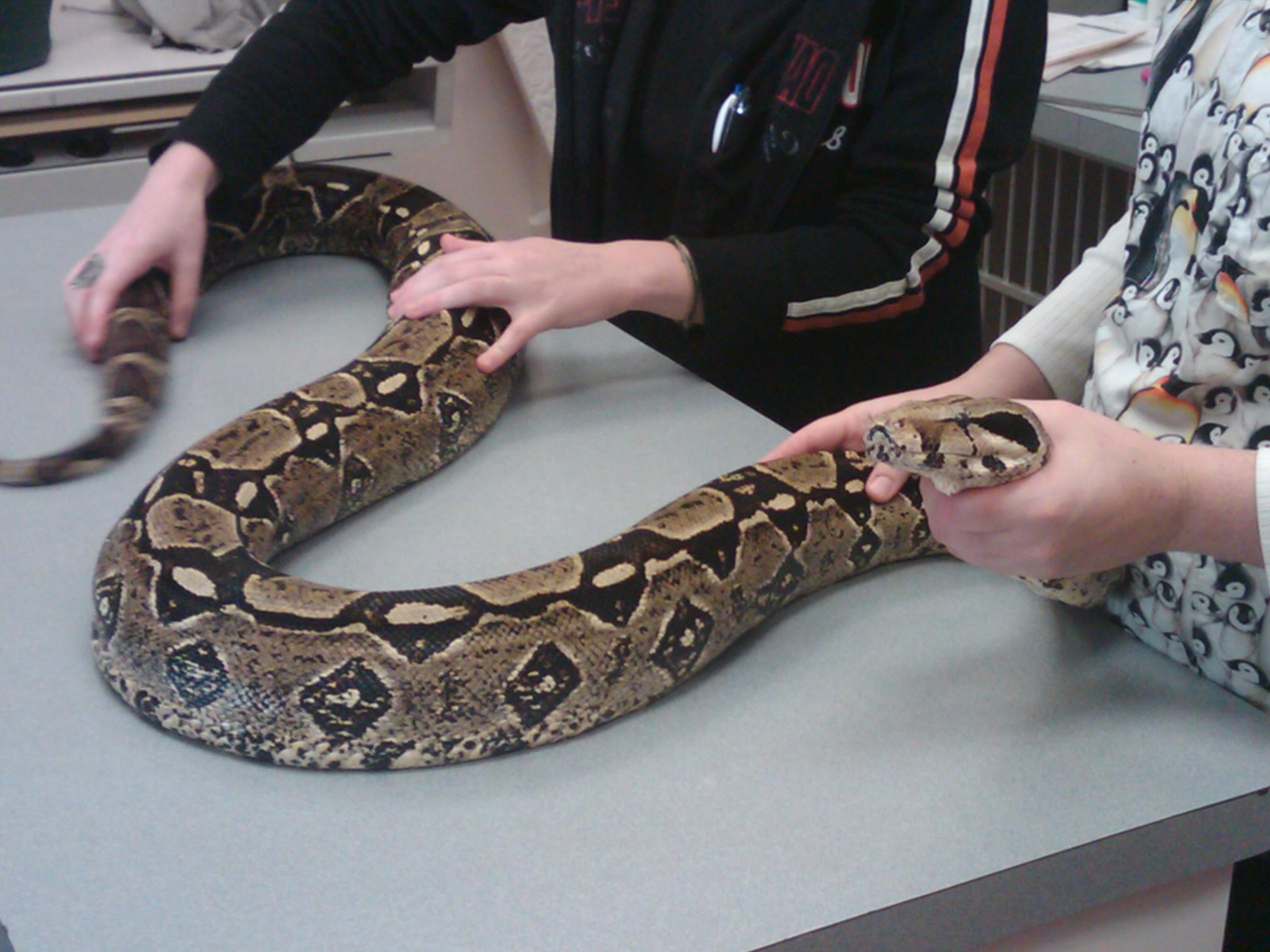
History: A 26-year-old male Boa Constrictor (Image 1) was presented for anorexia & weight loss over last 4 months. The clinical exam revealed good body condition and attitude. A caudal abdominal mass was palpable. Blood analysis revealed elevated total protein and calcium levels. Mild elevation of uric acid was also present.

Comments

Special thanks to: Todd Wolf DVM, DABVP & staff at Companion Animal Hospital, Parsippany, New Jersey, USA, for providing this patient for evaluation.

Clinical Differential Diagnosis

(Wolf DVM, DABVP): Caudal abdominal neoplasia of renal, reproductive, intestinal origin, abscess, granuloma, obstipation.

Sampling

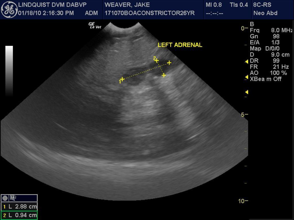
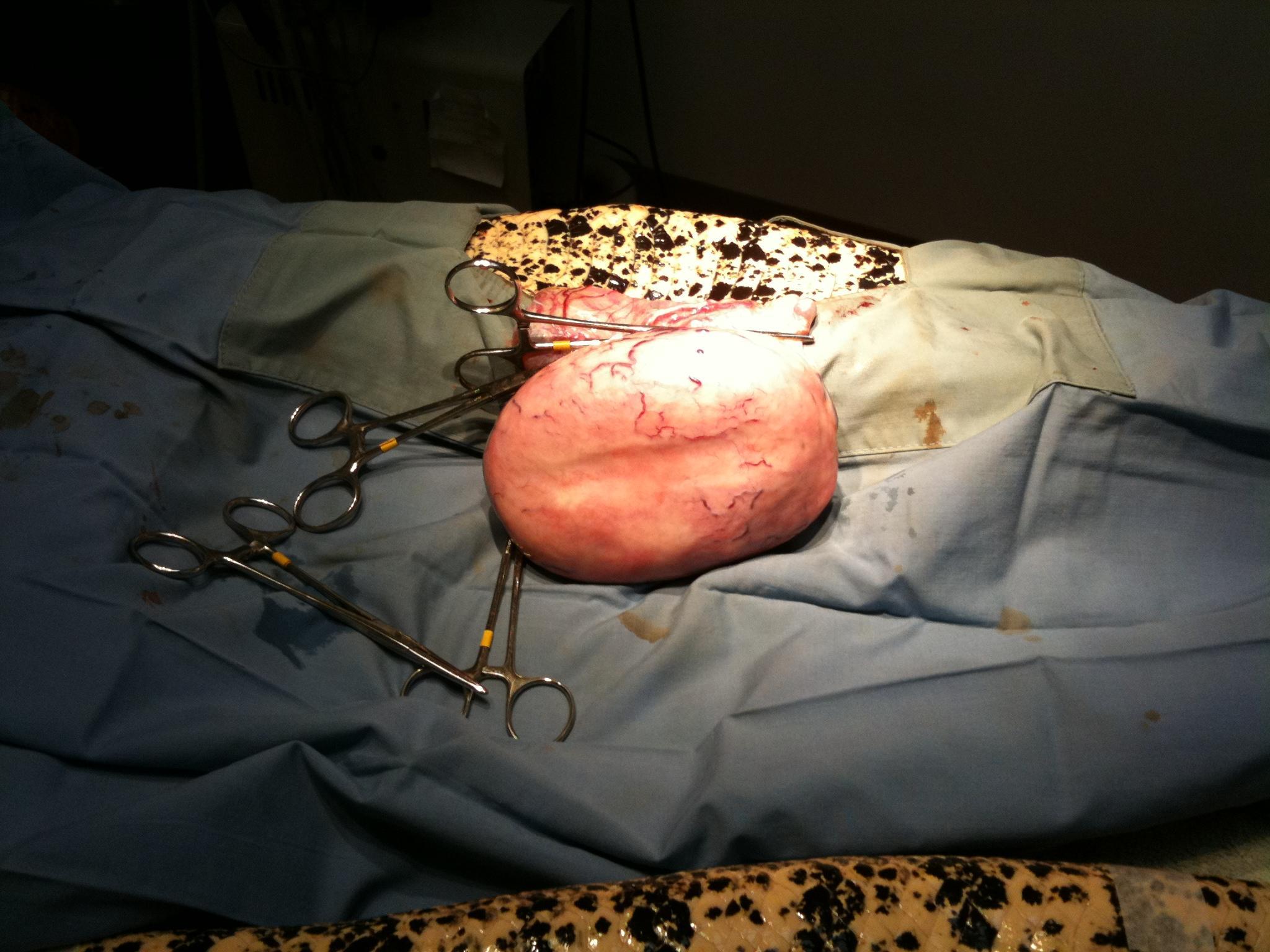
(Wolf DVM, DABVP): Laparotomy revealed a testicular mass that was successfully removed. Histopathology: Seminoma (Image 7)

Sonographic Differential Diagnosis

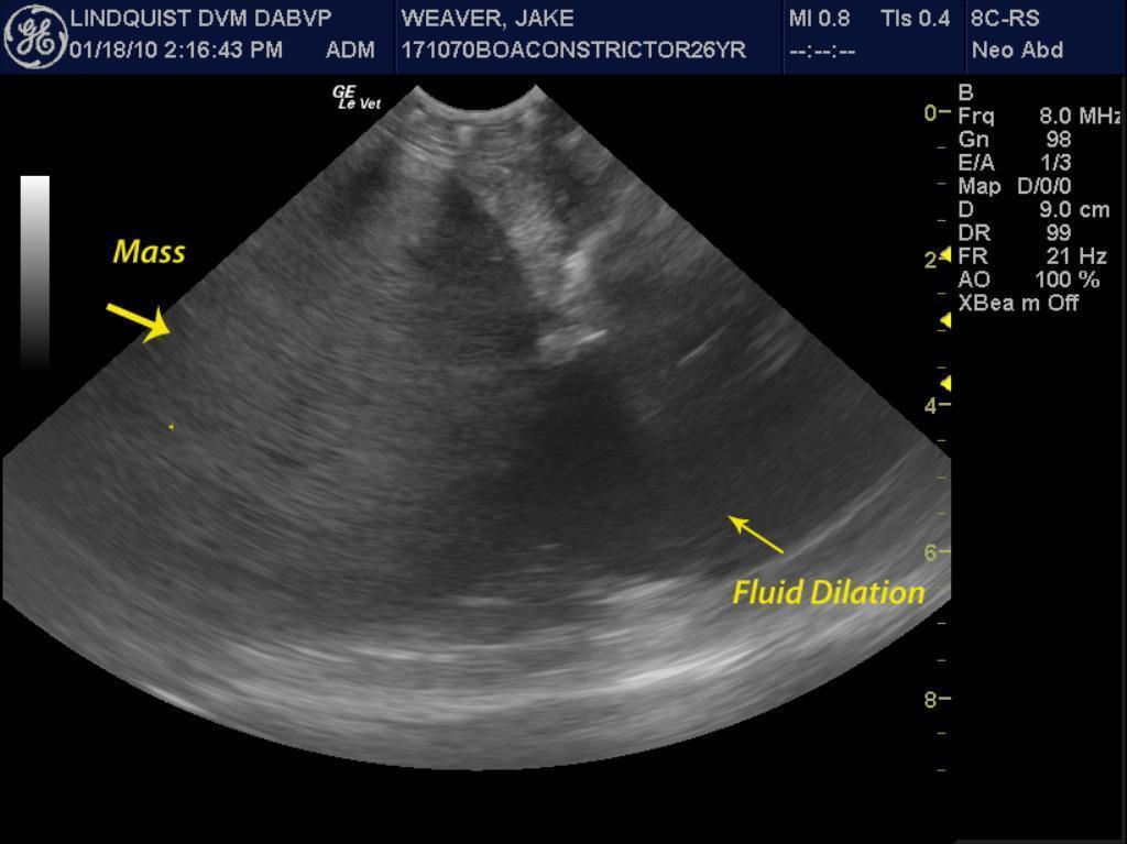
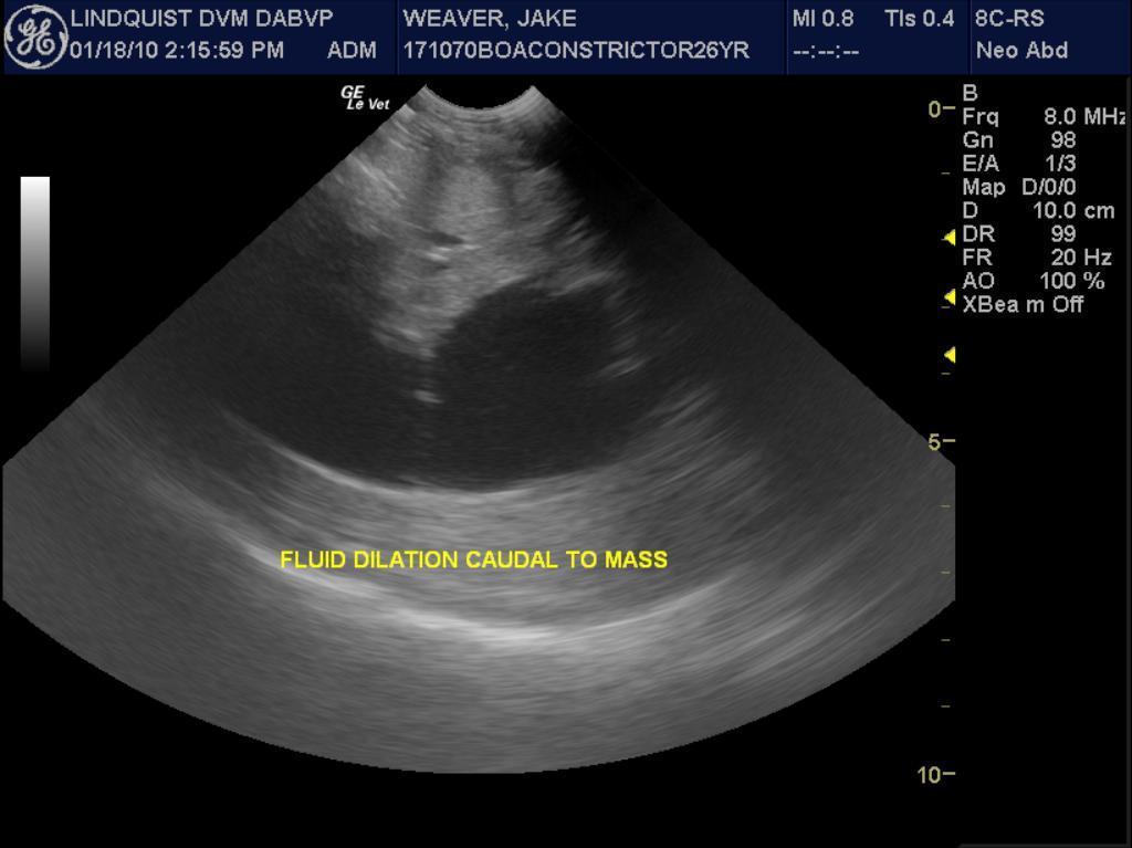
(Lindquist DMV, DABVP): Solitary mass deriving from reproductive or renal origin; likely surgically resectable.

Image Interpretation

(Wolf DVM, DABVP):
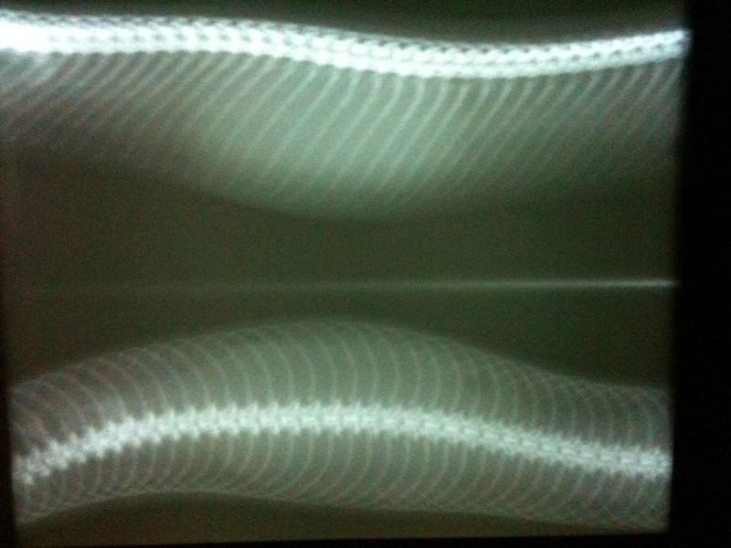
(Image 2) A homogeneous soft tissue mass is evident and rules out obstipation. Radiographic Differential: Caudal coelomic neoplasia/tumor, abscess, granuloma.

Outcome

The patient responded well postoperatively and was asymptomatic 6 weeks post-op and eating normally after 4 months of anorexia.

Patient Information

Age : 20 Years
Gender : Male, Intact
Species : Exotic
Status : Complete

Images

COM_1004_01COM_1004_02COM_1004_03COM_1004_04COM_1004_05COM_1004_06COM_1004_07

Skip to content