A 5-year-old male boxer dog was referred for evaluation of progressive lameness, poor appetite, and weight loss. Laboratory testing had been non-diagnostic.
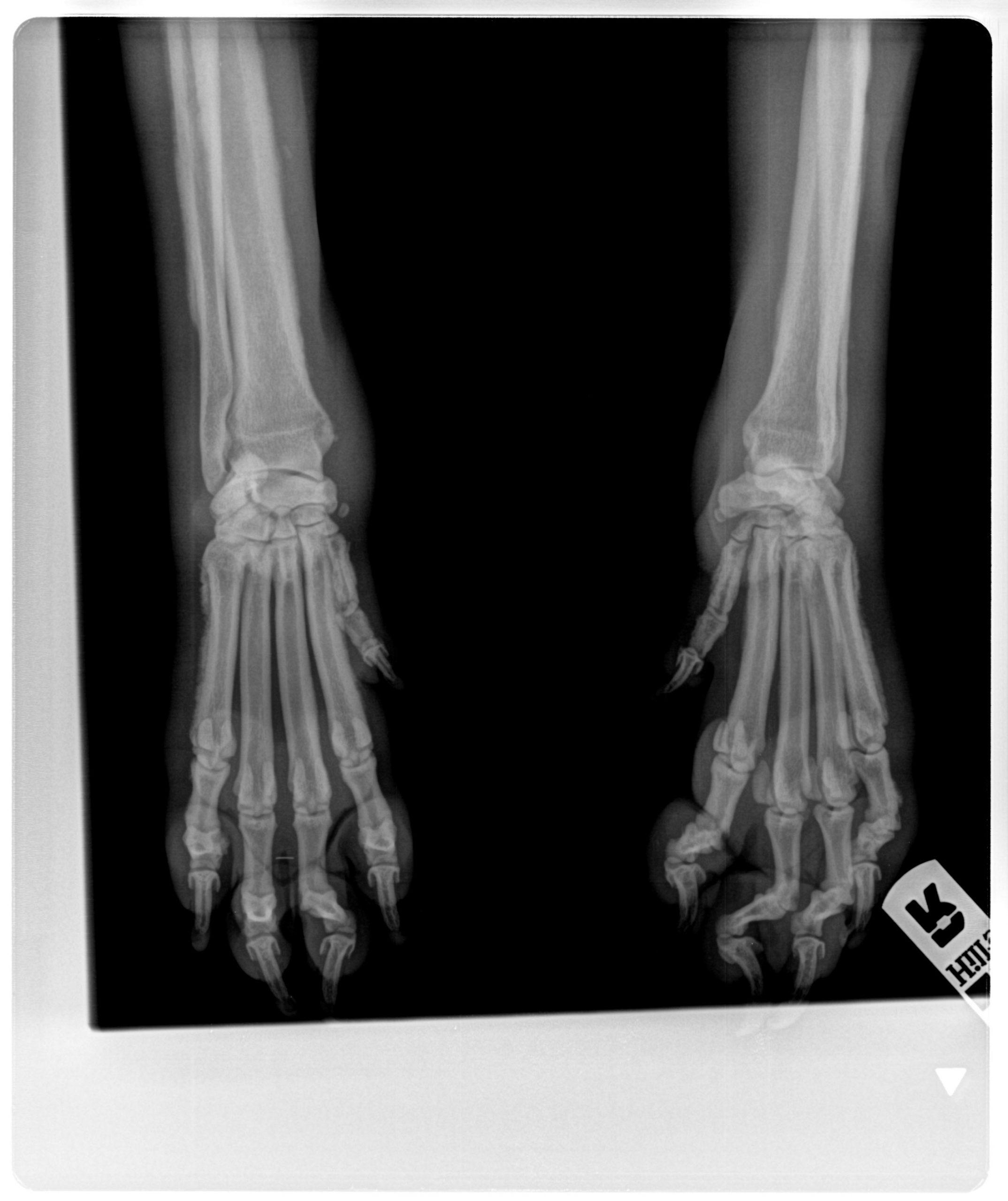
Abnormalities on clinical examination were weight loss, a stiff-choppy gait when walking, and firm swelling of the distal limbs with discomfort on palpation. Survey radiographs of the distal forelimbs showed a diffuse periosteal bone formation on the metacarpal bones and phalanges.
A 5-year-old male boxer dog was referred for evaluation of progressive lameness, poor appetite, and weight loss. Laboratory testing had been non-diagnostic.
Abnormalities on clinical examination were weight loss, a stiff-choppy gait when walking, and firm swelling of the distal limbs with discomfort on palpation. Survey radiographs of the distal forelimbs showed a diffuse periosteal bone formation on the metacarpal bones and phalanges.
Further diagnostics
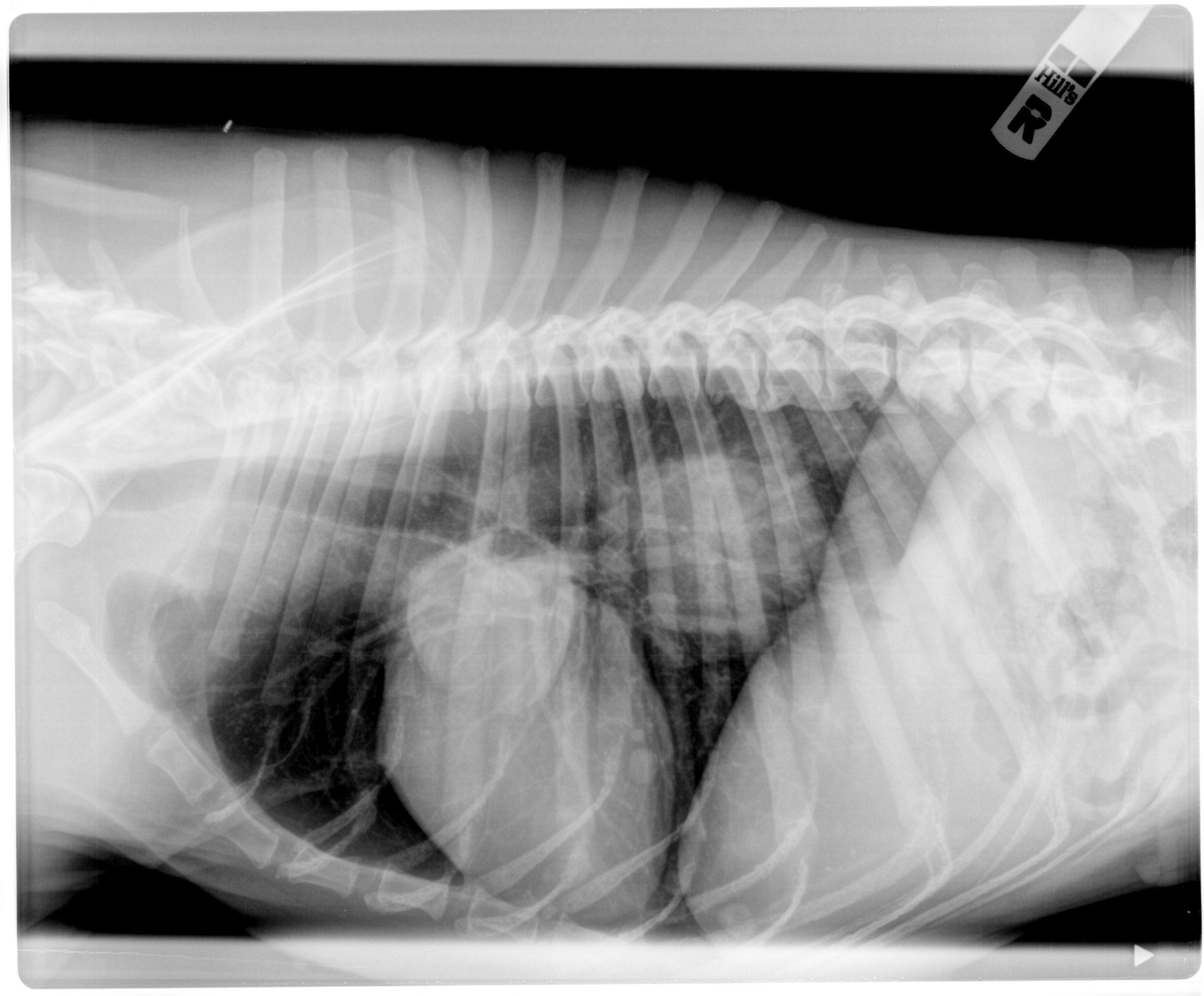
On survey radiographs of the thorax a caudal mediastinal mass (80 x 60 mm), a focal mass (60 x 60 mm) in the right cranial lung lobe, and smaller nodules in the caudal right lung lobes were evident. A mottled echogenic mass in the right thorax adjacent to the heart was present on thoracic ultrasonography. Fine needle aspirate cytology of the mass showed osteoid material, few neutrophils, and clumps of anaplastic osteoblasts.