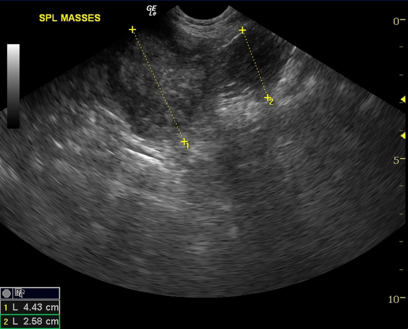
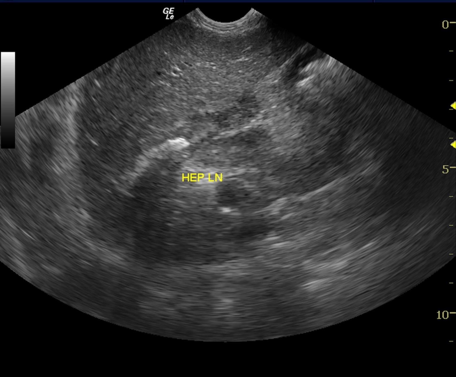
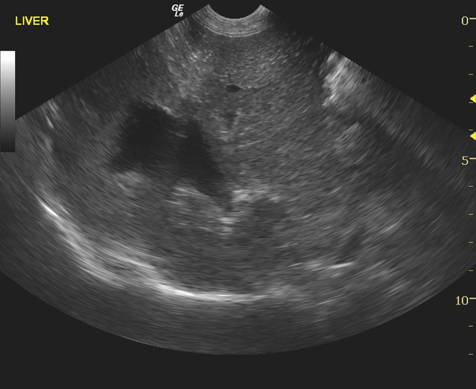
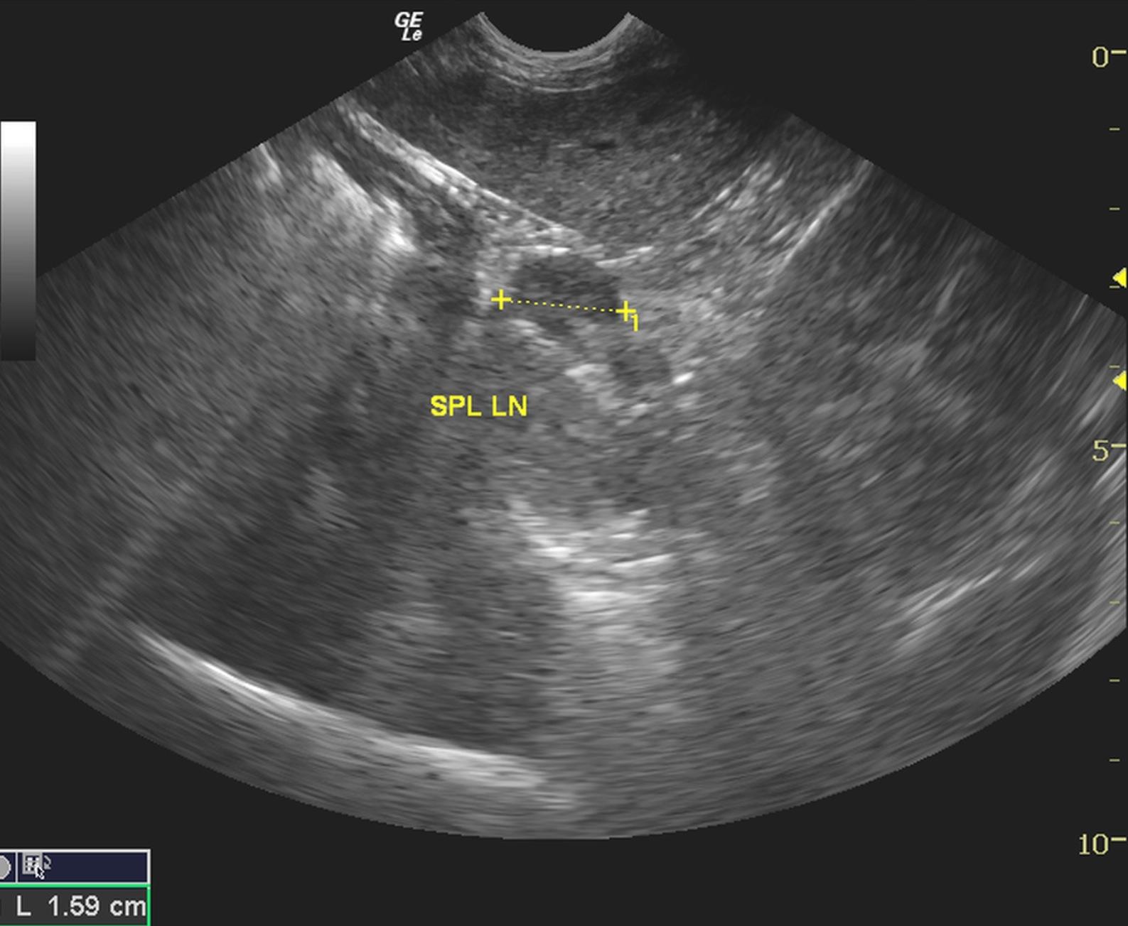
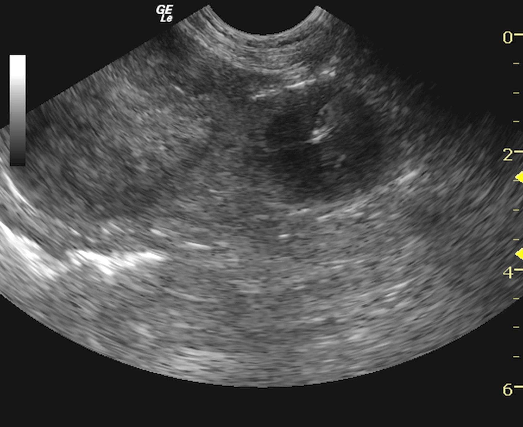
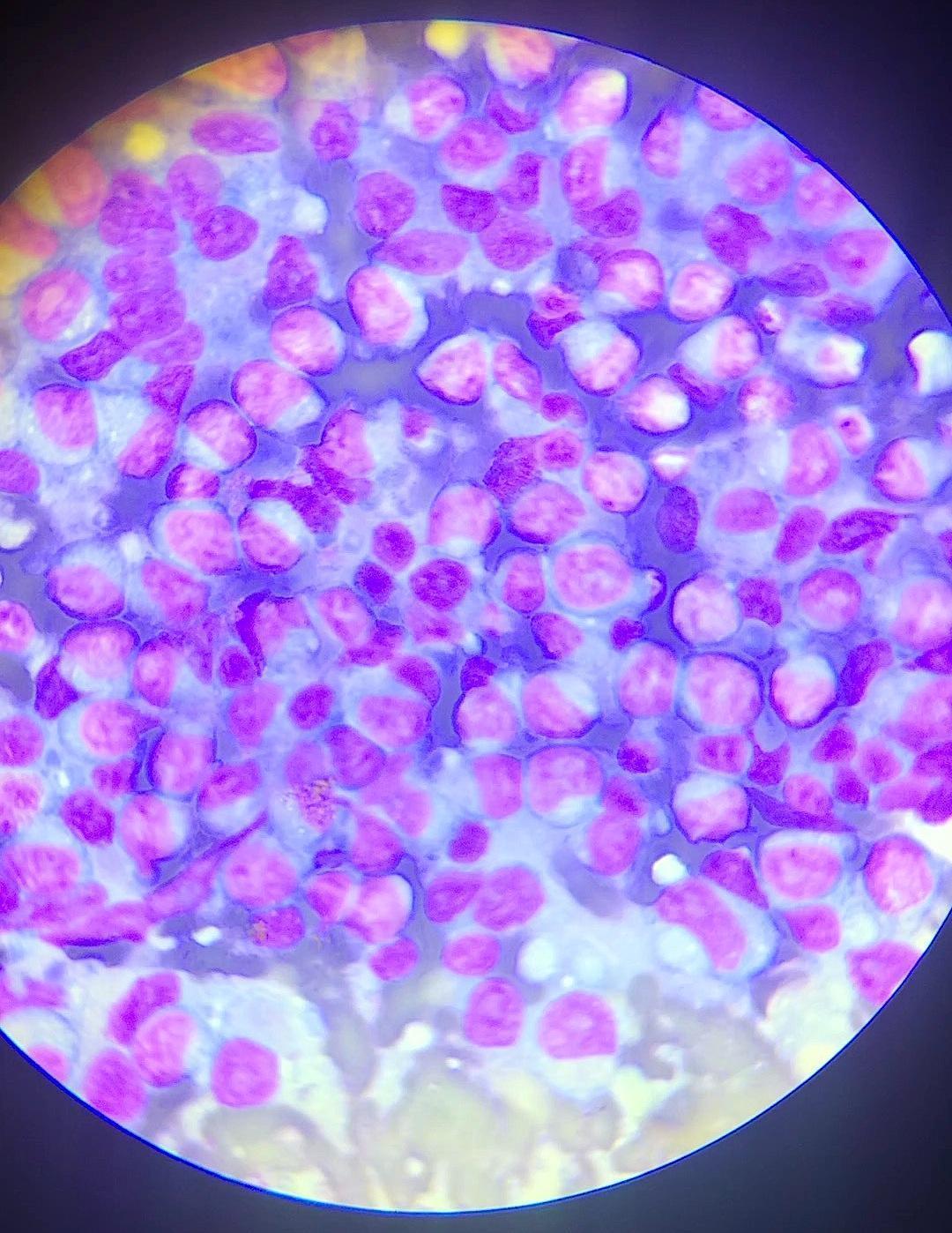
A 12-year-old MN Labrador Retriever mix was presented with chronic diarrhea +/- syncopal episodes.Low grade anemia was found on CBC with unremarkable blood chemistry findings.
A 12-year-old MN Labrador Retriever mix was presented with chronic diarrhea +/- syncopal episodes.Low grade anemia was found on CBC with unremarkable blood chemistry findings.