A 10-year-old FS Pit Bull terrier was presented for a long history of left front lameness.
A 10-year-old FS Pit Bull terrier was presented for a long history of left front lameness.
What is that forelimb limp all about? What’s going on in that shoulder? A probe on the joint with the proper protocol is all you need when our expert radiologist Dr. Nele Ondreka from the founding radiology department of orthopedic ultrasound in the 1970s, University of Giessen in Germany is interpreting the image set. If you haven’t seen what orthopedic ultrasound can do, you will never cut a knee, shoulder, or achilles again without a sonogram of the joint. Biceps tendinitis or tear? Synovial damage? Cruciate or meniscus partial or full tears? Joint DJD? It’s all in the report before you cut so you know what you are getting into first with orthopedic ultrasound. I know now that I would never cut a joint without scanning first. I wish I had this when I was a surgeon years ago. Our orthopedic ultrasound seminar is this month in beautiful Ho-ho-kus, NJ (30 min from EWR) with a couple of spots left for wet lab and many for lecture (GPs and surgeons to know applications of ortho US) and CE credits as well to fill your year-end CE needs
A 10-year-old FS Pit Bull terrier was presented for a long history of left front lameness.
A 10-year-old FS Pit Bull terrier was presented for a long history of left front lameness.
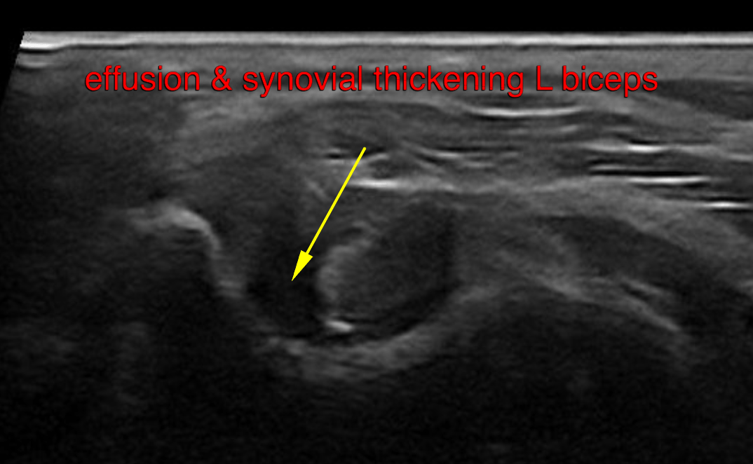
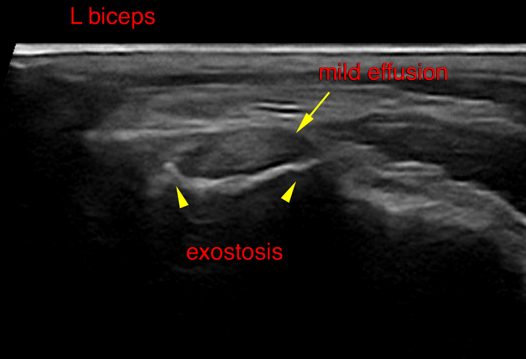
Left shoulder: Mild tendon sheath effusion and thickening of the synovial membrane is noted. The intertubercular groove presents a moderate exostosis encompassing the biceps tendon. Moderate osteophyte formations are noted at the periarticular margins of the shoulder joint.
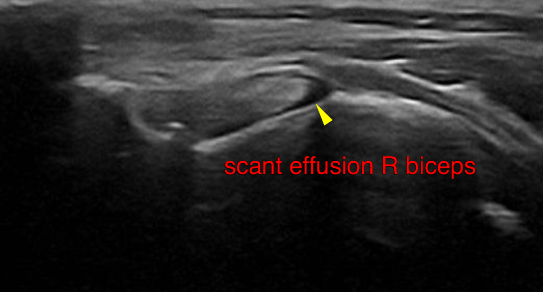
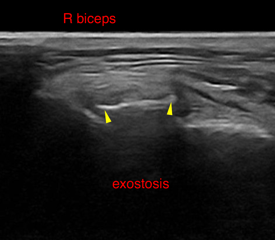
Right shoulder: Mild tendon sheath effusion is noted. Synovial thickening is not evident. The intertubercular groove presents a mild exostosis encompassing the biceps tendon. Moderate osteophyte formations are noted at the periarticular margins of the shoulder joint.
At this stage conservative management with PRP injection into the left shoulder joint (automatically permeates into the communicating biceps tendon), rest, systemic anti-inflammatory treatment and targeted physical therapy should be considered as a clinical trial for the lame left front limb. However, some patients with exostotic bone formation do not respond well to conservative management and may require arthroscopic revision with tenotomy even at relatively early stages of the disease. Consider proactive local PRP injection and physical therapy for the right front limb as well as it’s a bilateral disease in this patient and favoring of the left front limb may propel progression of the changes on the right side.