Intestinal Lymphoma in a 7-year-old FS Bullmastiff: Our Case Of the Month December 2016

December’s Case of the Month hits very close to home. Gabby, this beautiful 7-year-old bullmastiff, is the beloved pet of one of SonoPath’s SDEP Ultrasound Instructors Rebekah Jakum, CVT, ARDMS/RVT (and mobile sonographer for Pennsylvania Mobile Veterinary Ultrasound). Join us by following Gabby’s journey through the aggressive progression of intestinal lymphoma. All of the imaging and sampling was done utilizing SonoPath’s telemedicine and telecytology services resulting in exceptional turn around times to aid in diagnosis, expedite treatment options and support quality of life decisions.

History

A 7-year-old FS Bullmastiff was presented for acute diarrhea and decreased appetite.

A 7-year-old FS Bullmastiff was presented for acute diarrhea and decreased appetite.

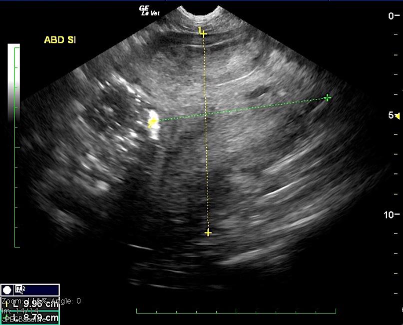
Sonographic summary: The initial ultrasound findings were: Part of the small intestine presents a 1:1 muscularis/mucosa ratio with decreased motility, mildly obscured wall layering and mild accumulation of chyme. Focal circumferential wall thickening of up to 7 mm with transmural loss of layering is seen in the ileocecal region and is associated with an incomplete obstructive pattern with segmental dilation of the intestine with chyme. The central abdominal mesentery presents a generalized increase in echogenicity with loss of the regular echoarchitecture emphasizing the lymph node and intestinal wall changes. Severe mesenteric lymphadenomegaly of up to 8 cm is noted. The lymph nodes are rounded with a pathologic increase of their short-to-long-axis ratio beyond 0.5. A significant mass effect on the intestine resulting in an incomplete obstructive pattern is noted. The margin to the intestinal wall is obscured. Scant anechoic peritoneal effusion is noted. Ultrasound guided fine needle aspirations were performed for further definition and confirmed a large cell lymphoma. The patient underwent chemotherapy and presented two months later due to extreme ADR since finishing the 2nd round of chemo (Adriamycin), anorexia with the exception of treats, and PU/PD. Subcutaneous lymphoma of a single mass had been detected via FNA 1 week prior, therafter multiple SQ masses developed.

The progression of pre-chemo, post chemo, and escape from remission sonographic images are sequenced below.

Comments

Thank you to Julie Holland, DVM of Littlestown Veterinary Hospital, Littlestown, PA and Ann Jeglum, VMD Veterinary Oncology Services and Research Center West Chester, PA for managing Gabby’s case.

DX

High-grade lymphoma

Sampling

Initial FNA of mesenteric lymph node found large cell lymphoma. Fine needle aspirate of a subcutaneous mass several weeks later was consistent with high-grade lymphoma. Submission of pleural fluid was also consistent with high-grade lymphoma.

Image Interpretation

Initially severely distorted lymph nodes were present with regional peritonitis and variable infiltrative intestinal pattern. After chemotherapy a significant reduction of the pathologic presentation was noted. Minor fluid accumulation was noted in the small intestine. The mesenteric lymph nodes were only slightly enlarged and measured 1.0 cm. They were mildly rounded. This is consistent with lymph nodes in remission. Escape form remiossion later occurred demonstrated by non cardiogenic pleural effusion noted in the thorax with variable lung consolidations. Cranial mediastinal lymph nodes were enlarged, distorted and hypoechoic measuring 2.0 x 2.5 cm.

Outcome

Unfortunately despite chemotherapy, supportive care, and tons of love, Gabby’s disease had progressed to the point where quality of life had declined and she was humanely euthanized.

Video

Patient Information

Patient Name : Gabby Jakum RJ's parent's dog šŸ™
Age : 7 Years
Gender : Female, Spayed
Species : Canine
Status : Complete
Code : 04_00562

Clinical Signs

  • “Not Doing Right”
  • Anorexia
  • Diarrhea
  • Vomiting

Images

gabby_jakuminitialuslnsi_gabby_2016_

Skip to content