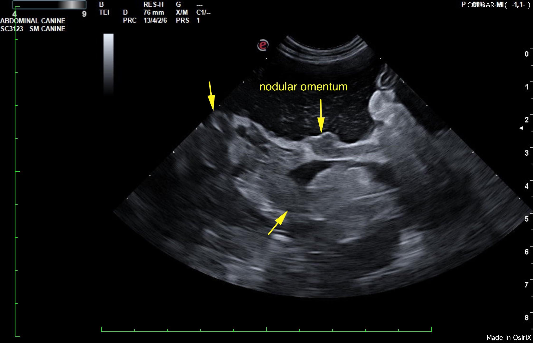
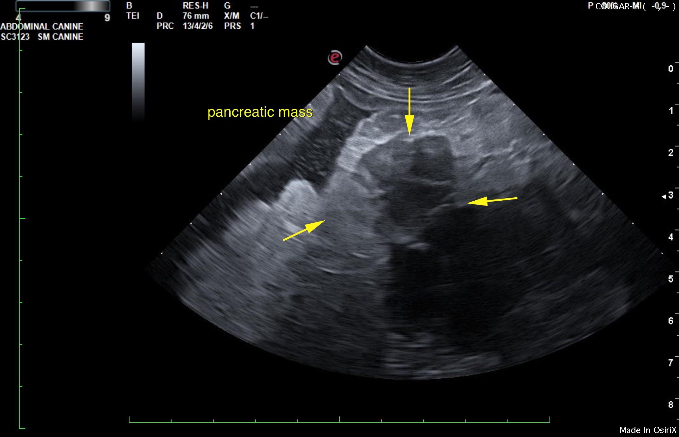
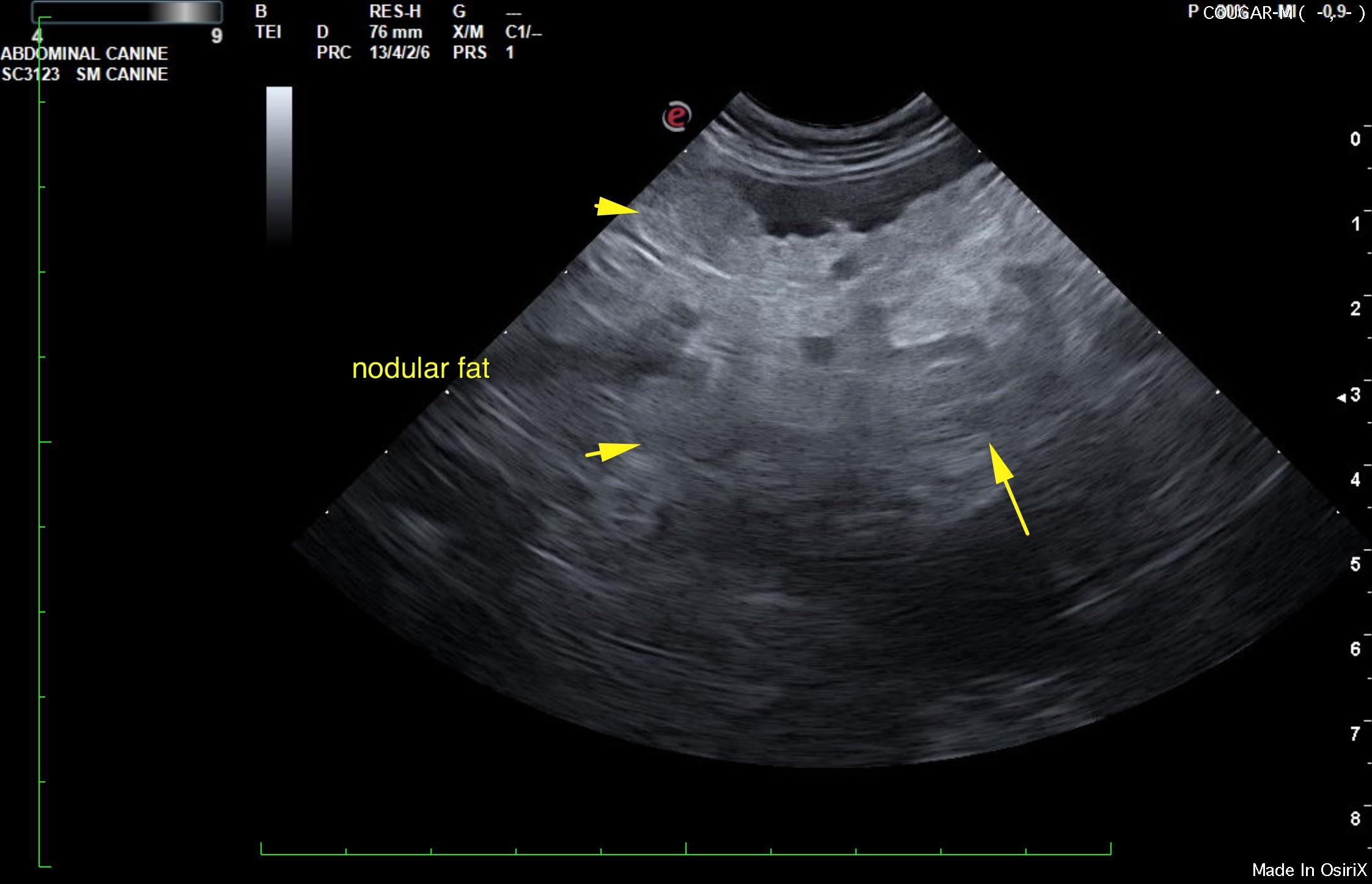
What’s that rough hair coat, abdominal swelling, and “ADR” elderly cat all about? Carcinomatosis, which is usually of the pancreatic origin, may be in play in these elderly pot-bellied kitties. If you are seeing ascites for no other reason (albumin normal, no hepatic vein dilation and nothing is ruptured) along with a “cotton candy” omental envelopment of the pancreas with hypoechoic, ill-defined nodules, then think of carcinomatosis as a possibility. Perform abdominocentesis and cytospin the fluid or FNA the hypoechoic areas for the answer. Occasionally, necrotizing pancreatitis can look like this, but when the carcinomatosis quacks like a duck, it’s usually a duck.
An 11-year-old, 15 lb, MN, DSH cat was presented for a hernia and decreased appetite. Indoor only cat since a kitten and FELV/FIV negative as a kitten. CBC and blood chemistry showed WBC 20.66, neutrophils 17.29, low normal HCT/RBC/hemoglobin, platelets 1229.
An 11-year-old, 15 lb, MN, DSH cat was presented for a hernia and decreased appetite. Indoor only cat since a kitten and FELV/FIV negative as a kitten. CBC and blood chemistry showed WBC 20.66, neutrophils 17.29, low normal HCT/RBC/hemoglobin, platelets 1229.
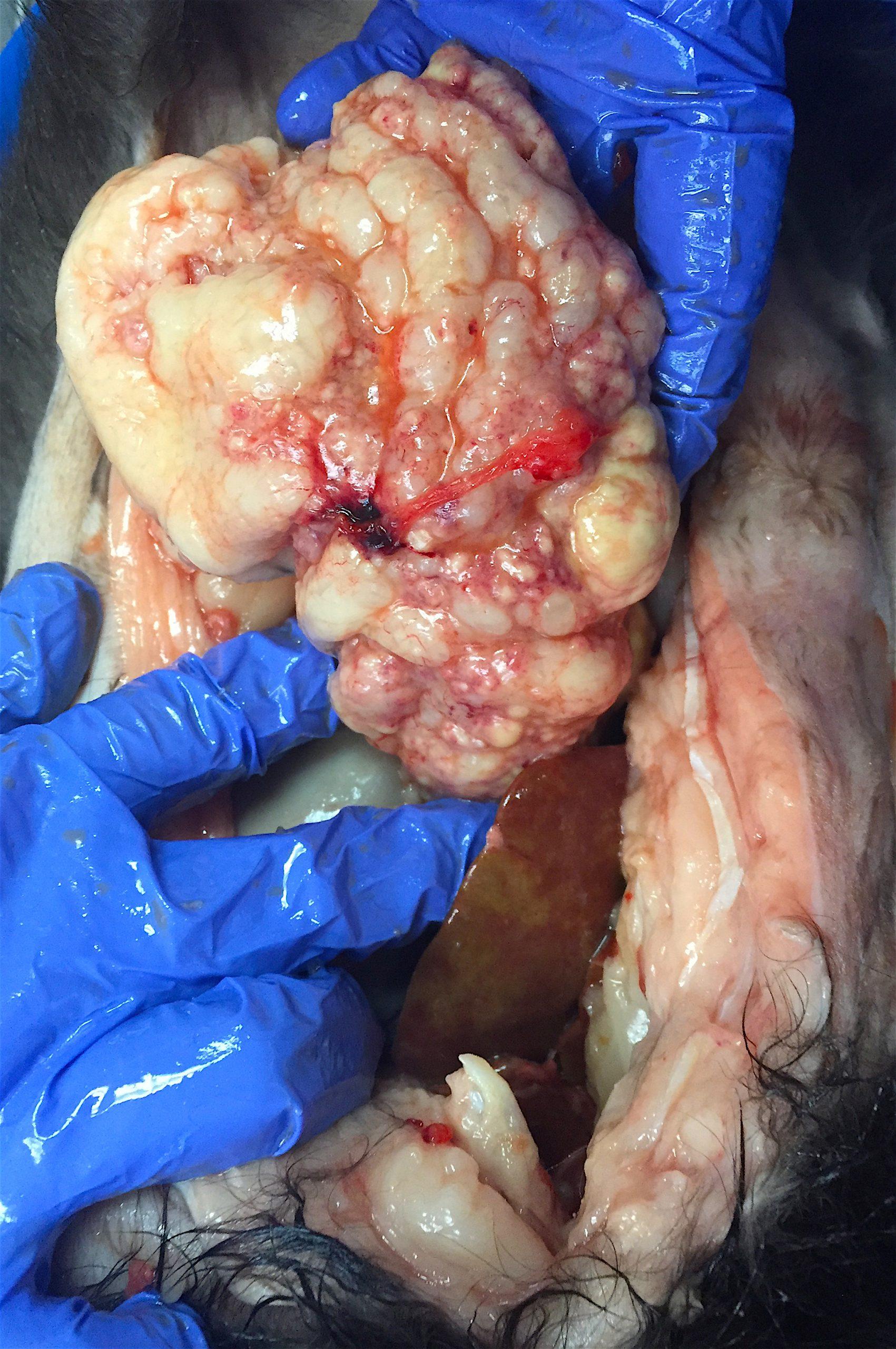
Pancreatic adenocarcinoma with marked necrosis and autolysis.
Ascites and nodular, irregular pancreas and omentum. Multiple biliary calculi; non-obstructive. Strong suspicion for pancreatic carcinomatosis or similar. Severe pancreatitis with necrosis possible, yet less likely.
The pancreas was enlarged, hypoechoic, and irregular. Nodular omental changes were noted. The pancreas was heterogenous in areas of hypoechogenicity and expansive irregular tissue strongly suggestive for underlyingpotential neoplasia or severe pancreatitis and pancreatic necrosis.