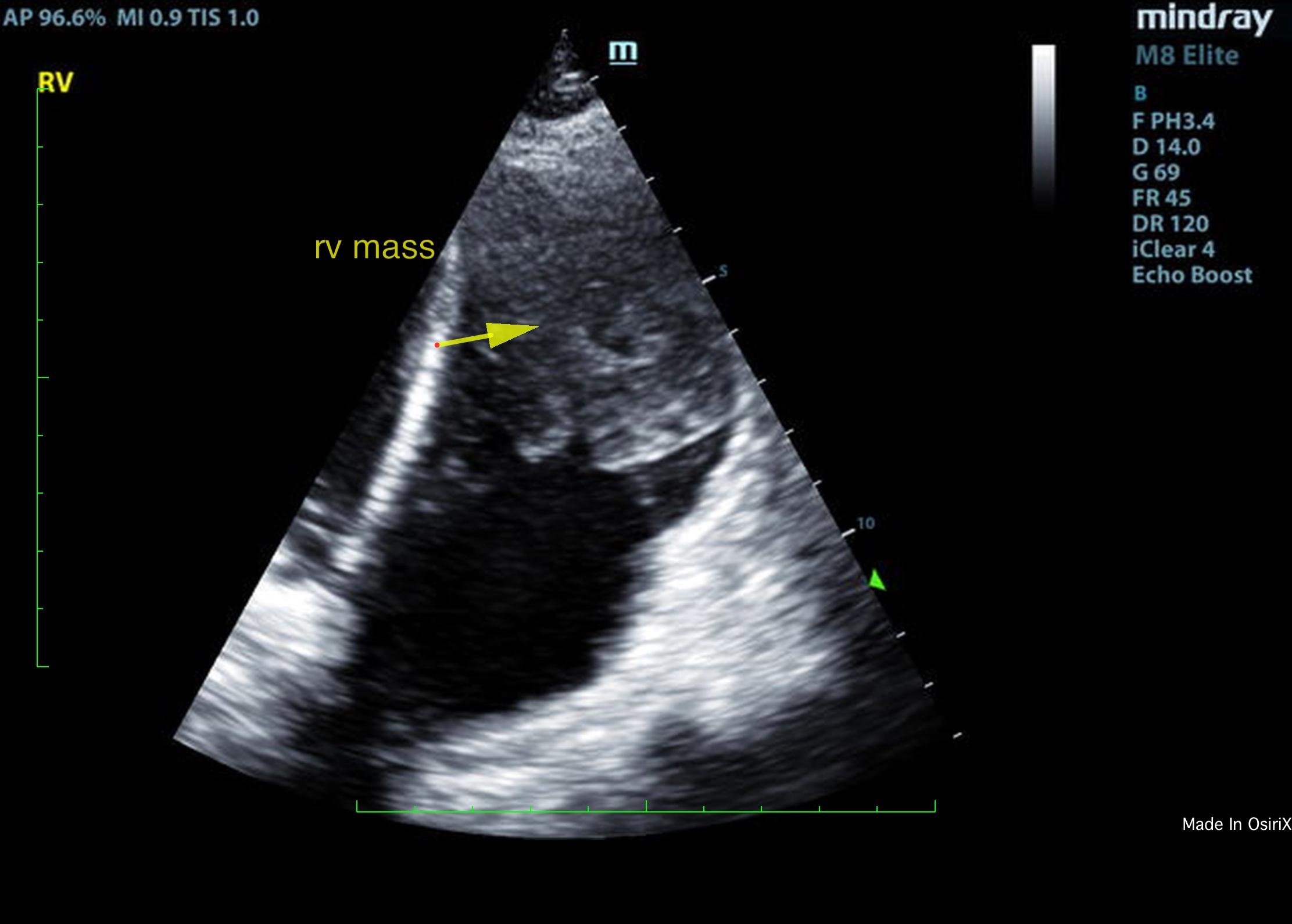
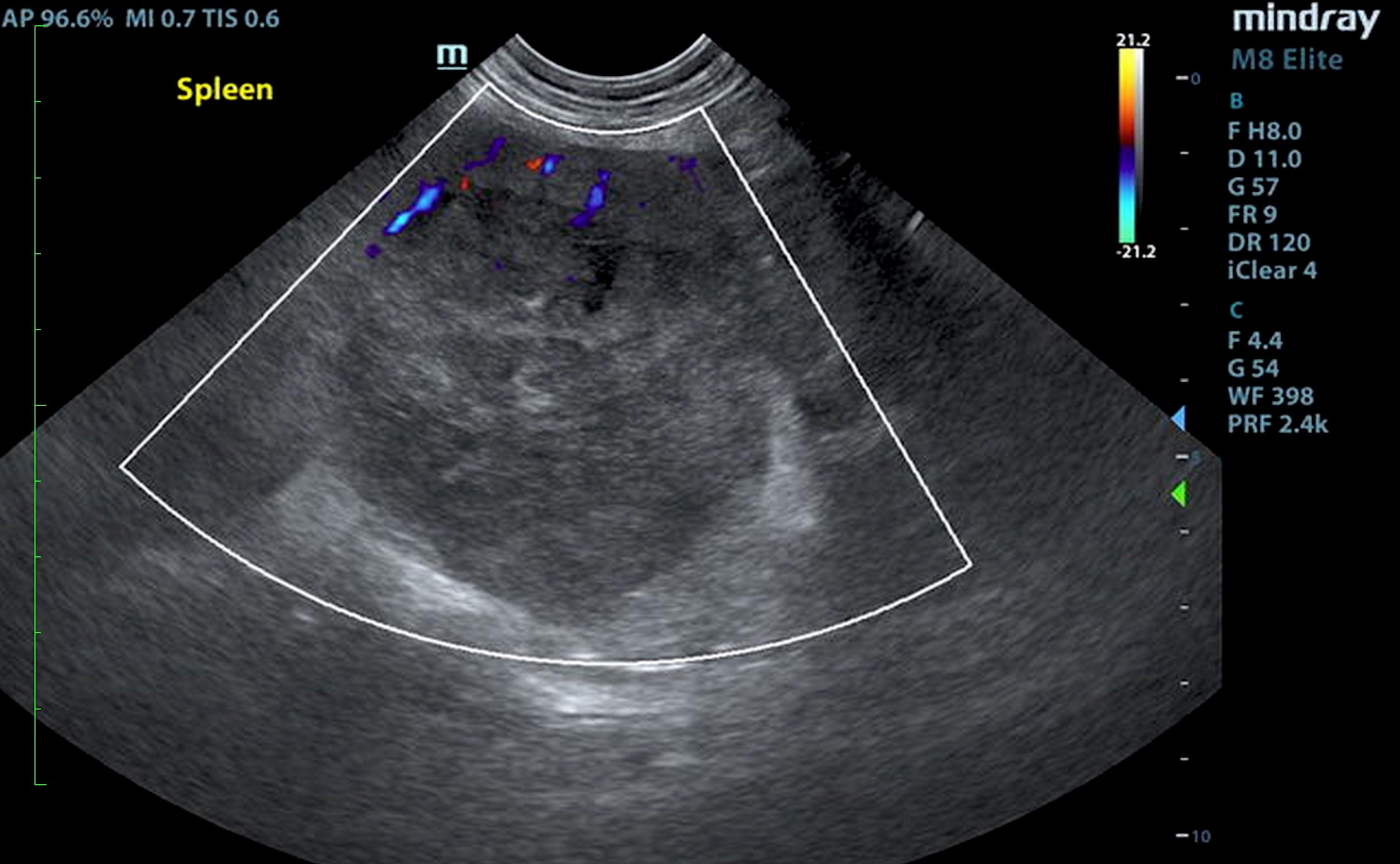
Expect to be surprised whenever you apply the probe – cardiac masses aren’t just for the right auricle and heart base. Consider this right ventricular alien found on a recheck of a mitral murmur, attached to the right ventricular septum and sneaking into the pulmonary outflow. Here an expansive splenic mass complicates the scenario, but there can be other sources of hemangiosarcoma type presentations. When considering potential oncological intervention, following the echo with a full abdominal scan is always best practice.
Thanks to Stephanie Pearce, RVT, RDCS with Intrapet Imaging for the imaging, Dr. Gabrielle Weber with Warm & Fuzzy Veterinary Center in Middle River, Maryland for the case management, and SonoPath Specialist Dr. Eric Lindquist, DMV, DABVP for the thorough interpretation of the cardiac mass and associated aortic and tricuspid insufficiencies on this older Doberman.
Patient presented for newly diagnosed grade 2/6 heart murmur, sudden weight loss, and newly diagnosed hypothyroidism. Previous history of random vocalization- unsure if from anxiety or discomfort.
Patient presented for newly diagnosed grade 2/6 heart murmur, sudden weight loss, and newly diagnosed hypothyroidism. Previous history of random vocalization- unsure if from anxiety or discomfort.
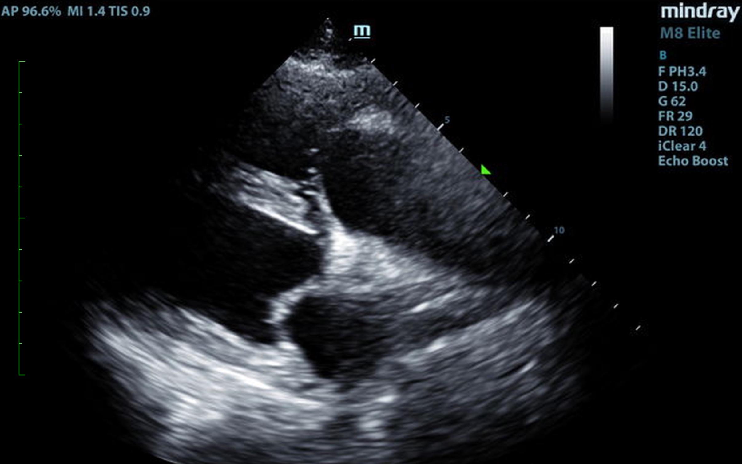
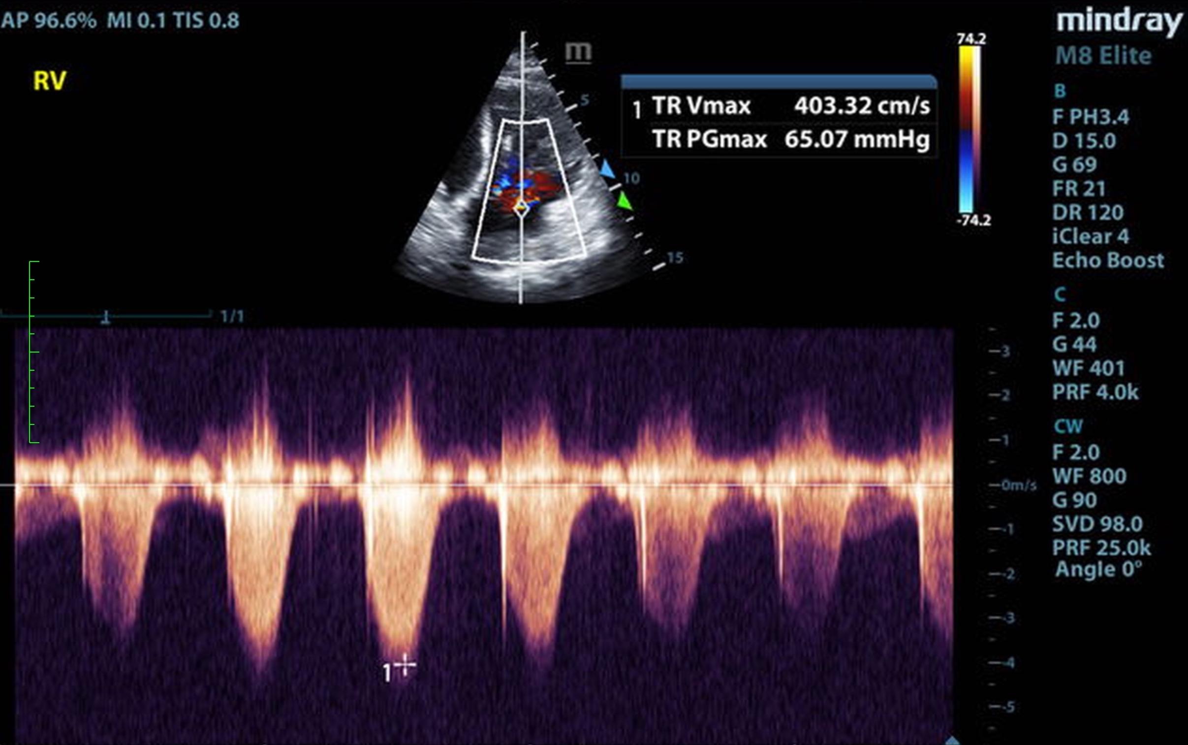
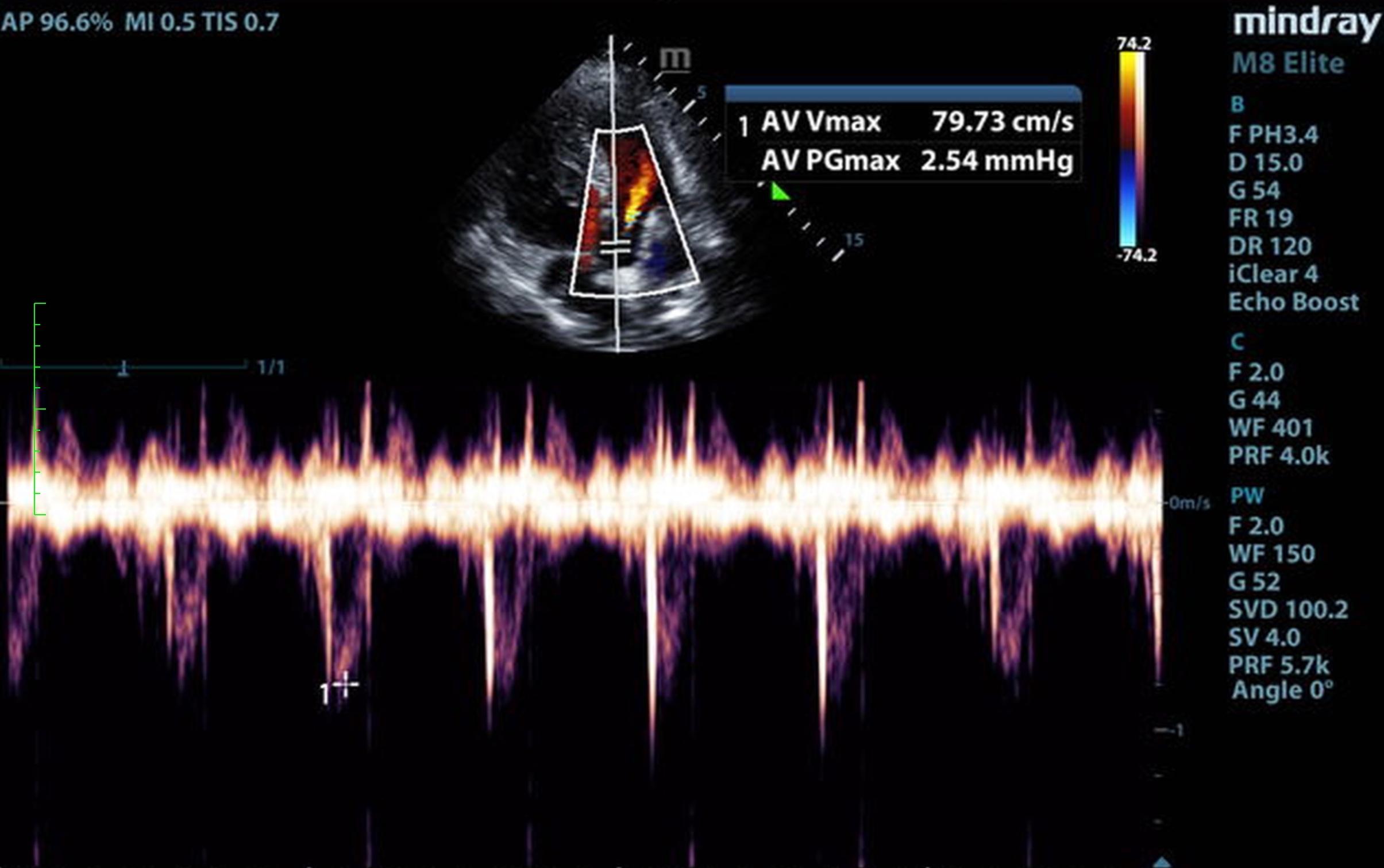
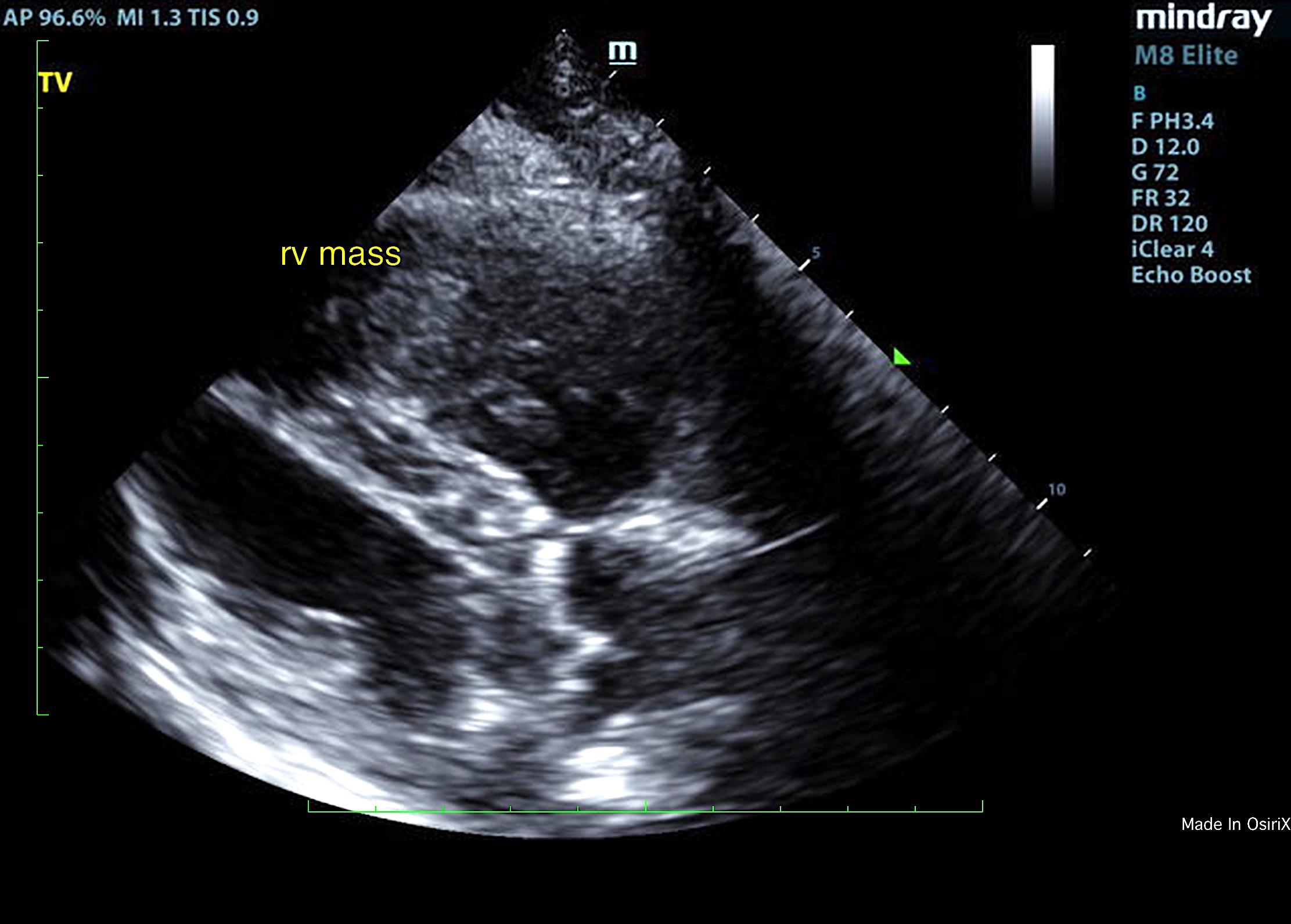
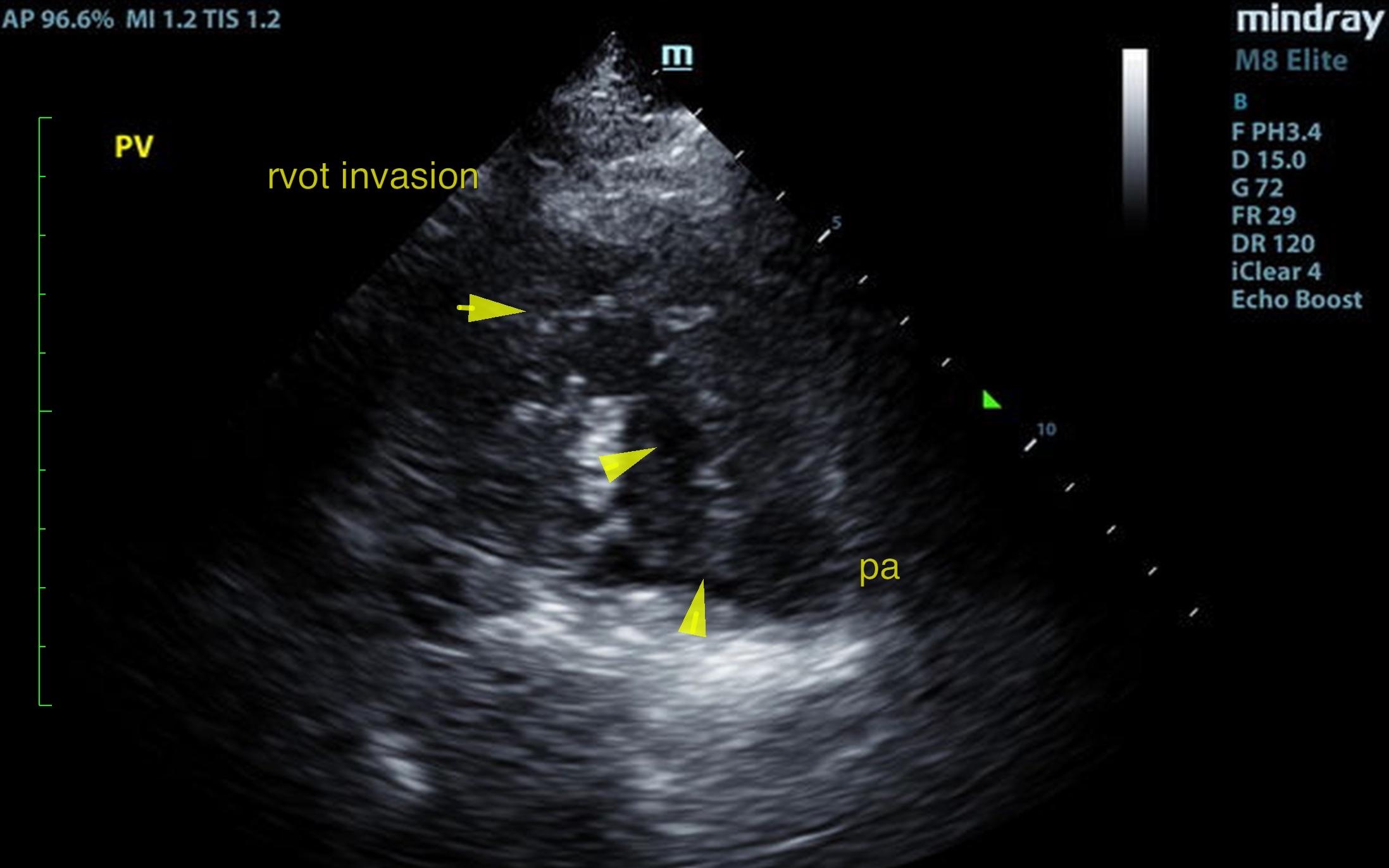
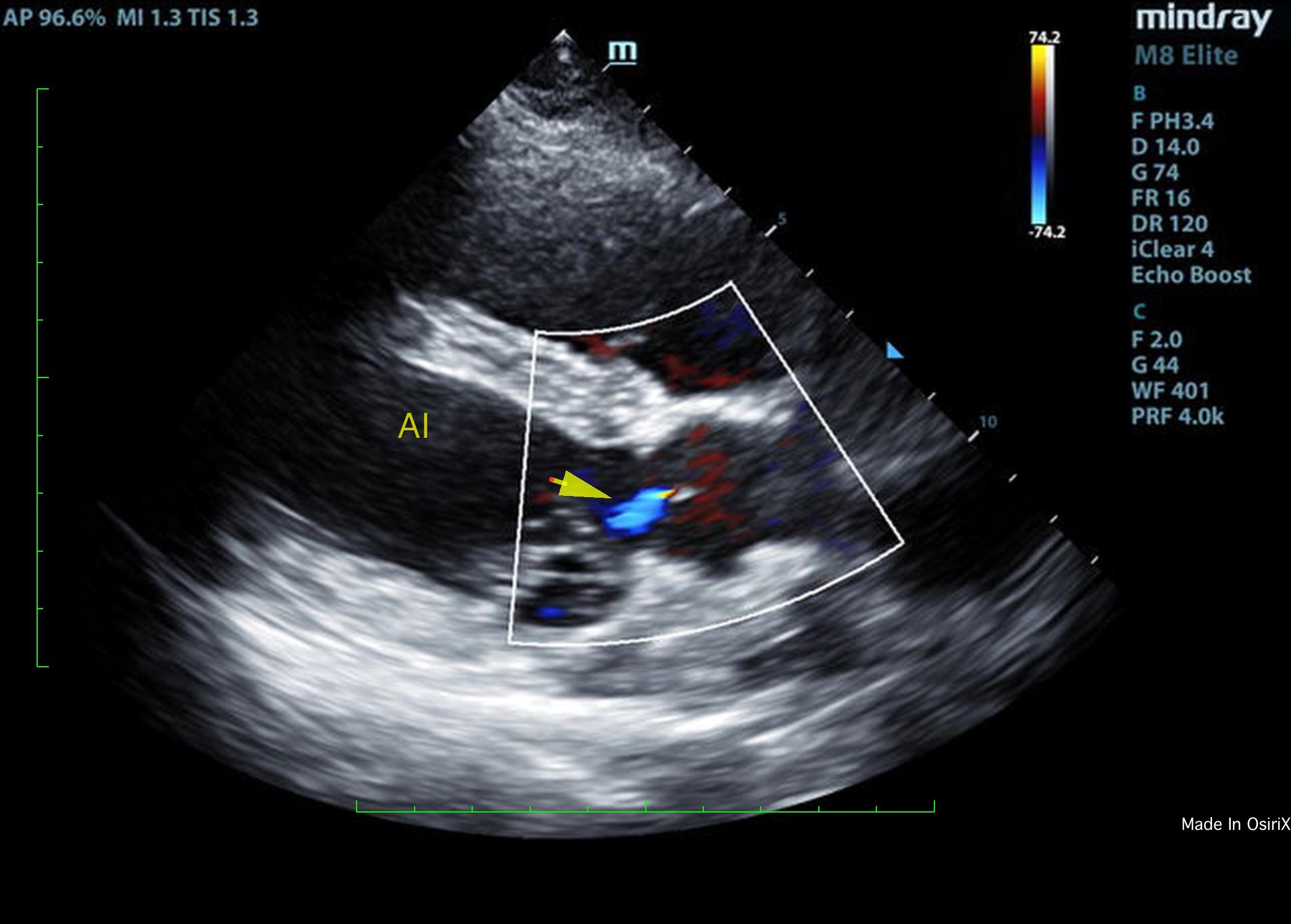
Right ventricular papillary mass obstructing the pulmonary outflow. Flattened ventricular septum. Secondary pulmonary hypertension. Concurrent aortic insufficiency. Splenic mass.
Flattened ventricular septum was noted as well. Aortic insufficiency was present on color flow Doppler. The right atrium was mildly enlarged with significant tricuspid insufficiency with velocities consistent with severe pulmonary hypertension. Right ventricular septum revealed a mixed, hypoechoic mass. Given the splenic pathology metastatic hemangiosarcoma is likely. The mass entered into the right ventricle and measured approximately 3.0 cm. No pericardial effusion was noted. However, the right ventricular mass entered into the pulmonary outflow as well. This created an obstructive flow pattern. There was mild volume overload noted in the right ventricle as well. Spleen – The spleen revealed a moderately complex, mixed hypoechoic expansive mass that measured approximately 6.0 cm.
Given the splenic mass combination with the right ventricular mass hemangiosarcoma or similar sarcoma is
likely. Chemotherapeutic trial can be considered in this patient to reduce mass volume with Doxorubicin or similar. This may also reduce the splenic mass. Oncological intervention is recommended. The patient was humanely euthanized due to quality of life issues.