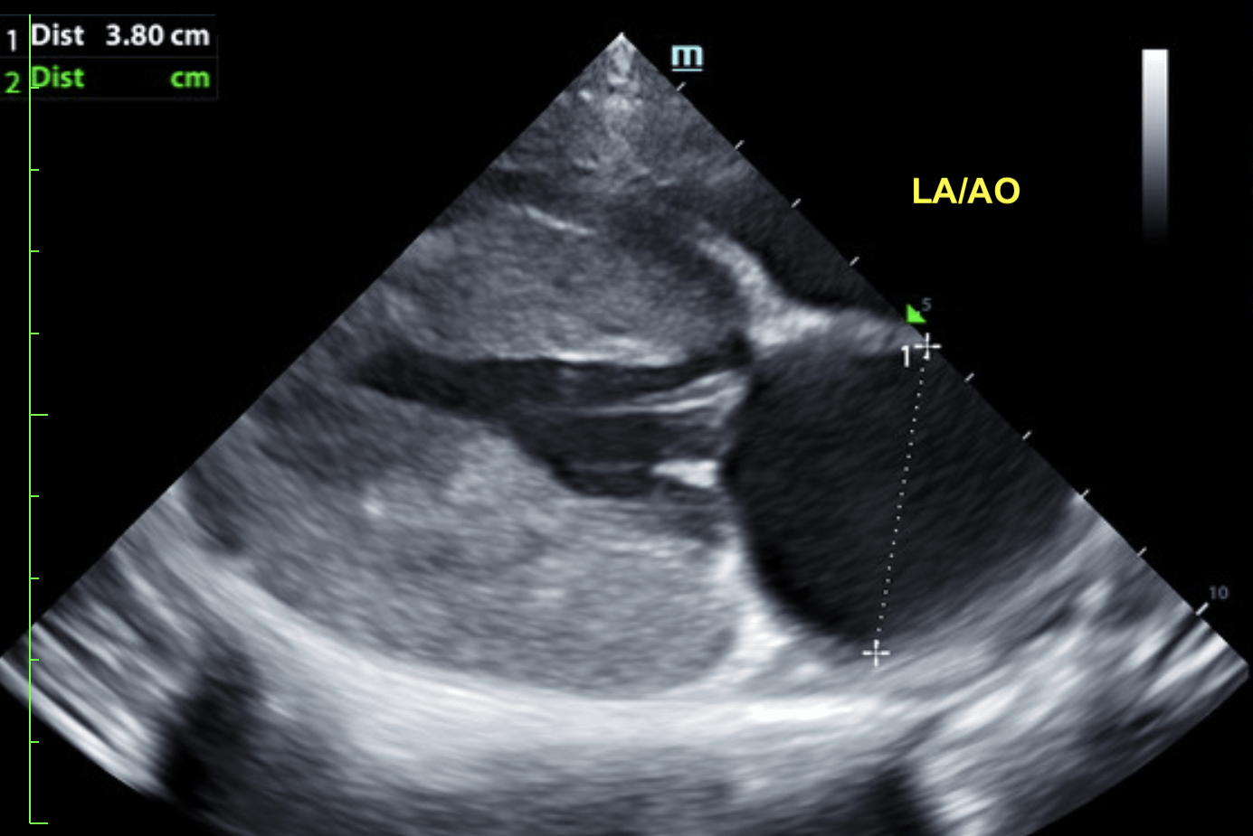
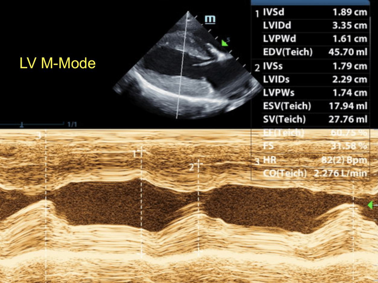
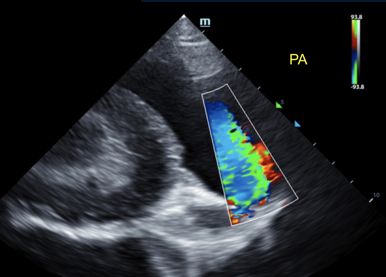
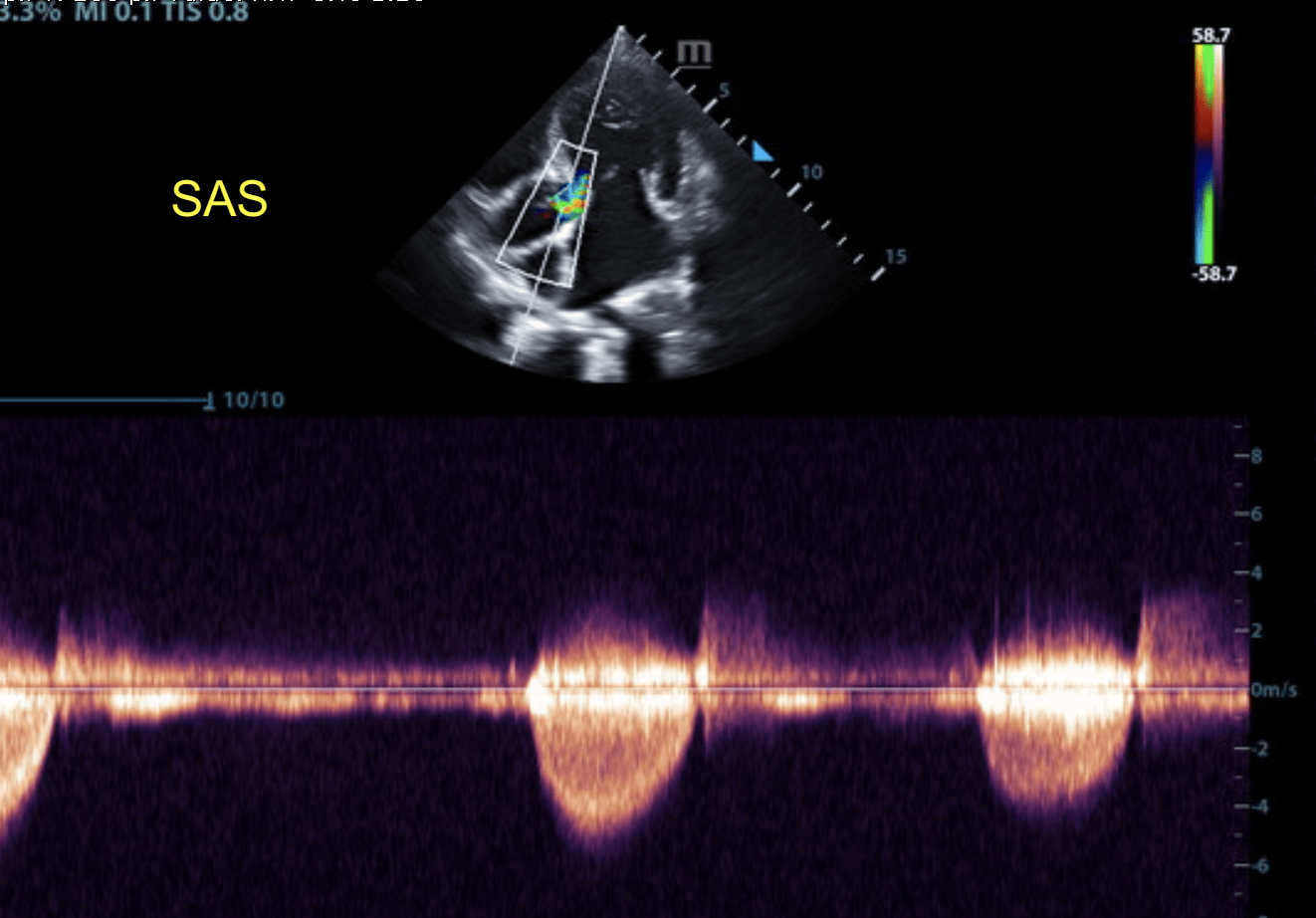
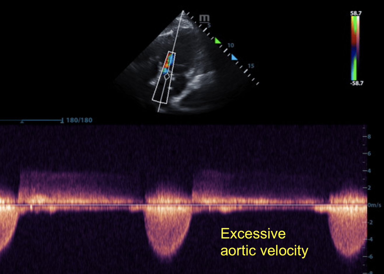
A 9-month-old intact male Mastiff mixed breed with history of being underweight presented at a referral facility for echocardiogram due to heart murmur.
A 9-month-old intact male Mastiff mixed breed with history of being underweight presented at a referral facility for echocardiogram due to heart murmur.