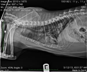
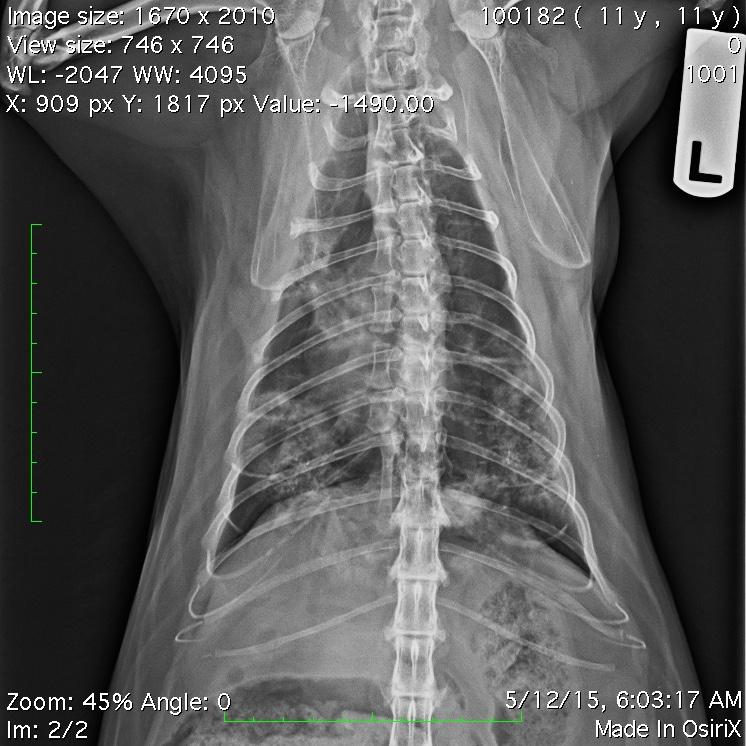
I was looking for some advise. I had a cat in today with Tachypnea with dry rales. I sent the X-rays to IDEXX for a radiology consult. They suggested I consider an ultrasound guided FNA of one of the nodular areas.
I have never done this before.
I would appreciate some advise including advise on restraint.
Do you sedate or place under general anesthesia?
Any info would be helpful.
I would also like to know if I can send DICOM x-rays for Dr O’brien to read as a telemedicine consult.
I could send .jpg’s- but I you really can’t do much with the images.
Thanks


10 responses to “FNA Thoracic Mass”
Sedation vs anesthesia very
Sedation vs anesthesia very dependant on the patient. In calm/relaxed animals there is often no need for sedation and easily done under US guidance. FNA done the same way as you would aspirate an abominal mass/liver/etc. Looking at the radiographs, seems that you would be able to visualize the mass in the right caudal lung field with US.
Randy take a look at the
Randy take a look at the arrows to approach the lesions closest to the body wall. Have a tech compress the lung under sedation like this technique in resources: http://sonopath.com/resources/interventional-procedures/lindquist-compression
and look for consolidations similar to what is present in this case in the archive: http://sonopath.com/members/case-studies/cases/pulmonary-carcinoma-diagnosed-lung-fna-12-year-old-fs-kerry-blue-terrier
measure out well how far you need to put the needle into the consolidated lung so you don’t end up doing a CVC or cardiac blood sample 🙂 but you should be fine away form anything dangerous given the caudal aspect of the lung lesions.
Thank you EL. The last
Thank you EL. The last suggestion cleared things up for me. I will let you know how this
turns out.
Thank you EL. The last
Thank you EL. The last suggestion cleared things up for me. I will let you know how this
turns out.
Shortly after I posted the
Shortly after I posted the x-rays I brought Takota back in for a FNA of the lungs. I must admit I never could identify a solid mass therefore cytology was not submitted- although I did do an aspirate where I felt there was pathology. Nothing worth sending in.I started Takota on Prednisone and a shot of convenia. He is doing a bit better. RRR is in the mid to high 20’s/minute- but he is still wheezing.
I did offer the owners a referral to an internal medicicine specialist- but they said they would not go that far.
Here are the follow up x-rays one week later.
I see some improvement- but I still think neoplasia is a possiblity here.
Any possiblity this could be chornic airway disease with fibrosis and calcification. Almost hoping for something similar to COPD.
Hey Dr O’brien- if you ever check in I would also value your imput.
Thanks
Neoplasia typically doesnt
Neoplasia typically doesnt get better in a week but that event doesnt rule it out. Unfortunately now the right caudal lesion is tougher to stick because its resolving. Its a tough first stick if you haven’t done lung fna in the past. The key is to count the ribs, left side recumbency after sedation and look for the consolidation compressing the lung with your scanning hand til you get a window to the non aerated consolidation. Look at my instruction on lung compression technique.
https://sonopath.com/resources/interventional-procedures/lindquist-compression
Here are some applicable cases:
http://sonopath.com/members/case-studies/search?text=lung+mass+fna&species=All