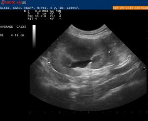
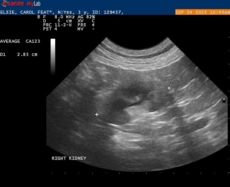
Elsie is a 4 year old female spayed DSH. She came in for an exam pre-dental. Horrible periodontal disease with many teeth missing. This cat came from a rescue.
Results of pre-dental bloo work:
UA: SG 1.016, pH 6.0, Dipstick: 3+ hematuria. Sed: 1+ protein, 10-15 RBC, WBC: 0-2/HPF, Bacteria: none seen, rare epi cell. The hemauria may have been iatrogenic from the cystocentesis. The bladder looked normal on ultrasound.
Profile: BUN: 57, Creat: 2.1, SDMA: 22 (<13)
T4: 2.2
CBC: WNL
Leuk/FIV: negative
Blood pressures around 140- normal
X-rays: mildly enlarge L kidney/small R kidney with a very tiny nephrolith noted in the R kidney.
I had concern about the hematuria, Stage 2 CRF normotensive and ? proteinuric
The abdominal ultrasound was totally normal excpt for the kidneys. I would like an opinion on the kidneys.
To me it appears there is poor cortical/medulary definition and gross dilation of the renal pelvices. The R kidny is much smaller than the L kindey. Could this be a congenital problem or is the small R kidney secondary to obstruction from a ureterolith?


3 responses to “Abnormal Kidneys – Feline”
Bilateral pyelonephritis
Bilateral pyelonephritis patterns on both and the right is the more progressed version. Sometimes the renal pelvices are strictured from stone passage. USG Pyelocentesis 25g on the LK and culture/decompress and tx aggressive pyelonephritis 3-5 days in house then long term and rescan weekly. Look for hypertension as well in near future since not there yet. Nice image set!
Thanks EL.
I have never
Thanks EL.
I have never actually done a pyelocenteis. Can you describe the technique again?
I use a 25g x 1.5. inch
I use a 25g x 1.5. inch needle and press the body wall down to the kidney as much as possible and pass into the renal pelvis at the shortest distance i can create manually from body wall to pelvis avoiding the vessels of course. Be sure not hypertensive first because hypertensive kidneys tend to bleed but with a 25g its usually not an issue just apply direct pressure for 30 sec after.Attached is what a typical sample may look like to culture but varies.
Viktor Szatmari wrote it up in javma a while back:
Szatmari V, Osi Z and Manczur F. Ultrasound-guided percutaneous drainage for treatment of pyonephrosis in two dogs. J Am Vet Med Assoc; 218(11):1796-1799, 2001.