Hello,
This is my third simmilar case and I thought that I should see what experts are thinking….
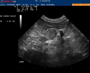
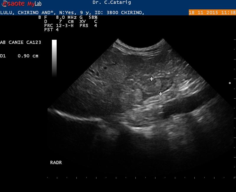
This is a 8 years old F/S dachshund that presented with moderate increse Liver enzymes. No clinical signs of PU/PD or symptoms for Pancreatitis. Both adrenal glands are enlarged, with “nodules” at the caudal pole and calcification/ s. Right limb of the pancreas is hyperechoic .
My questions :
1. Calcifications in the Adrenal gland are not suspicious of neoplasia, right?
Hello,
This is my third simmilar case and I thought that I should see what experts are thinking….
This is a 8 years old F/S dachshund that presented with moderate increse Liver enzymes. No clinical signs of PU/PD or symptoms for Pancreatitis. Both adrenal glands are enlarged, with “nodules” at the caudal pole and calcification/ s. Right limb of the pancreas is hyperechoic .
My questions :
1. Calcifications in the Adrenal gland are not suspicious of neoplasia, right?
2. If this dog doesn’t have symptoms of Cushings then I shouldn’t treat only monitor ( No Pu/PD USG 1.035)?
3.Is there any correlation between cushings disease and Hyperechic pancreas ( possible chronic pancreatitis) in dogs that are not currently diabetic or showing symptoms of pancreatitis; incidental finding ?
3 responses to “Adrenal Nodules, calcification and hyperechoic pancreas”
From Atlas of Small Animal
From Atlas of Small Animal Ultrasondography (penninck & Anjou; Blackwell Publishing 2008 Chapter 12 John Graham):
“The differential diagnosis for adrenal nodules and masses includes cortical adenoma, cortical adenocarcinoma, pheochromocytoma, metastasis, and hyperlasia. Cortical tumors may or may not be functional, and there may be no signs of hyperadrenocorticism. The sonographic appearance of these lesions is nonspecific. Clinical signs of pheochromocytomas are often vague, nonspecific, or intermittent, and their sonogrpahic features are nonspecific. Nodules and masses vary in size and echogenicity. However, if the nodules exceeds 2 cm, benign or malignant neoplasia is more likely, and if the lesion exceeds 4 cm, malignant neoplasia is more likely. However, both benign neoplasms and hperplasia may produce quite large lesions. The most reliable sign of malignancy is evidence of invasion of local tissues and structures, which can occur with adrneocortical carcinomas and pheochromocytomas.
Literataure references left out. See chapter in the book for specific references.
I hope this helps
These look like adenomas but
These look like adenomas but I pay more attention to them if systemic hypertension is present and of course if cushings is present…and always rescan in a month to check any growth. Adenoca and pheo can do this early and want to remove them if any growth and before phrenic vein invasion occurs if they are growing. If not growing then likely adenoma.
The hyperechoic pancreatic changes are consistent with amyloid or fibrosis… I see this in yorkies a lot and after history of pancreatitis.
Nice images!
Thank you both.
Thank you both.