- 2-3 year old stray mixed breed mn dog with recently diagnosed Canine Transmissible Veneral Tumor
- He has mulitple growths on his penis and a bloody prepucial discharge
- The ultrasound is part of a met check to see if the dog is suitable for adoption (by a veterinarian of course)
- Vincristine tx was started the day before this scan
- Abdominal US shows a mildly enlarged prostate (this dog was recently neutered)
- No enlarged lymph nodes, splenic or liver mets are seen
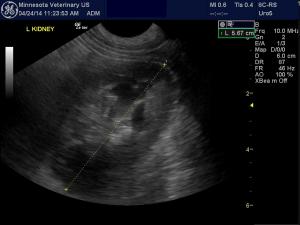
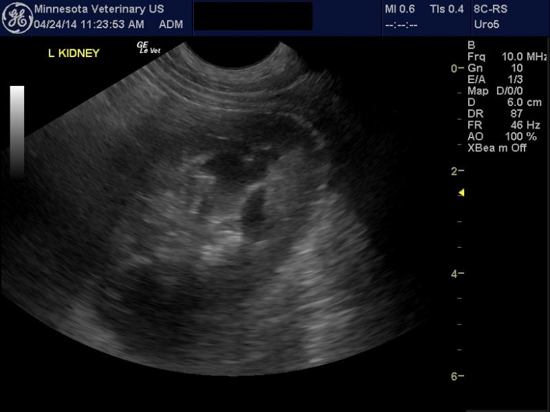
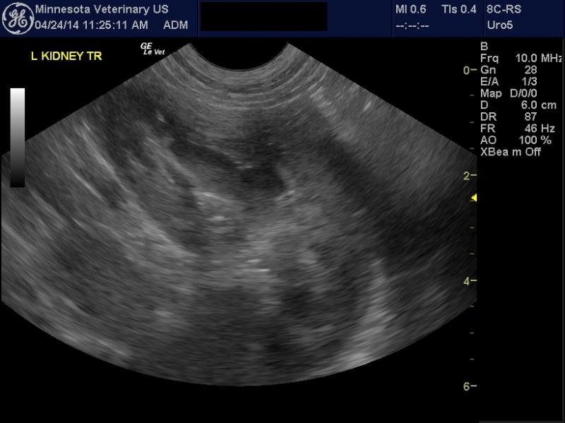
- The kidneys look “different” to me-Instead of a smooth renal cortex with a sharp delineation to a hypoechoic medulla, I am seeing lots more nodular “structure” and echogenic patches in the renal cortex and medulla.
- Are these battle scars from his life as a stray? Is this normal variation (I usually scan old dogs with loss of detail). I want to be sure there is no evidence of CTVT metastases, especially with the bloody discharge.
- I am unable to upload an avi clip as the files are too big.
8 responses to “Canine Transmissible Venereal Tumor”
The contour looks smooth. The
The contour looks smooth. The cm junction is a bit nebulous but may be the angle or the settings. The CM ratio is 1:1. They dont look bad to me and if no urinary sediment or prtoeinuria they are likely normal /stable maybe age related. Video may show more of what you are seeing. Usually when there are cortical changes infarcts and such some level of capsular deviation/retration occurs, even minimally and I don’t see that here.
It would very most unusual
It would very most unusual for a TVT only to metastasis to the kidneys. Spread to the kidneys is report but it is part of disseminated spead to various organs. Consider looking at a urinalysis and renal parameters on serum biochemistry to get a better idea of what the animal’s renal function is.
Thank you Dr. Lobetti. Renal
Thank you Dr. Lobetti. Renal parameters were normal. I have now successfully converted the videos to mp4 format and uploaded them.
Thank you Dr. Lobetti. Renal
Thank you Dr. Lobetti. Renal parameters were normal. I have now successfully converted the videos to mp4 format and uploaded them.
Still look clean to me
Still look clean to me
Still look clean to me
Still look clean to me