– 12 year old FS labradoodle presented for anorexia and distended abdomen
– pericaridal effusion, cardiac tamponade and ascites or hemoabdomen, large splenic mass and nodular omentum on ultrasound
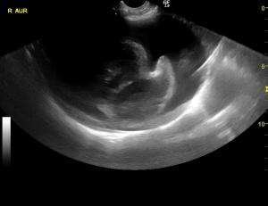
– just want to confrim that I am looking at a cardiac mass here – the actual right auricular appendage looks clean but I do see an irregualr hyperechoic tissue density when I scan between the left cranial long-axis right aurical and LVOT views.
– also suspicious region at the right parasternal heart base
– 12 year old FS labradoodle presented for anorexia and distended abdomen
– pericaridal effusion, cardiac tamponade and ascites or hemoabdomen, large splenic mass and nodular omentum on ultrasound
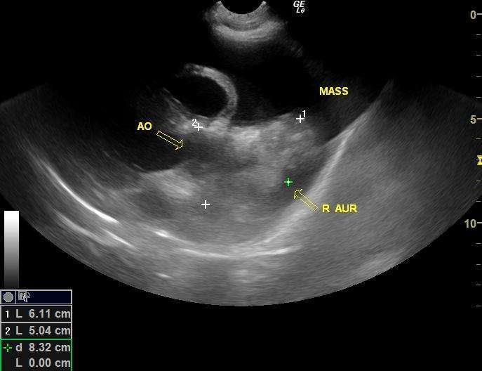
– just want to confrim that I am looking at a cardiac mass here – the actual right auricular appendage looks clean but I do see an irregualr hyperechoic tissue density when I scan between the left cranial long-axis right aurical and LVOT views.
– also suspicious region at the right parasternal heart base
3 responses to “Cardiac hemangiosarcoma”
yes I would call that a mass
yes I would call that a mass given the punctate hypoechoic ill defined changes within the muscular part of the atrium and invasion into the heart base. Clots attach within the pericardial space and dont invade like this but this is of course observational and its not like I get to histopath these guys at all.
Maybe Peter can chime in on this discussion as its a good one.
In my opinion what you see on
In my opinion what you see on the short axis view is not the same like what you see on the long axis view. On short axis the left ventricle fades in and out.This can give you a mass-like impression . I see this frequently when I review images (and scan the dogs myself afterwards). What you see on the long axis is – in my opinion – a mass. Hemangiosarcomas are usually seated on the right auricle – but not always. They can as well be attached to the heart base or the left atrium. Since you have a mass on the spleen, HAS is very likely.
PEter
Thank-you EL and Peter
With
Thank-you EL and Peter
With concurrent splenic lesions, this was a no-brainer but if they weren’t there I would have been second-guessing myself on this one. Happy Holidays!
Jacquie