A 2-year-old female Doberman was presented for evaluation of anorexia, renal failure, and vomiting. Azotemia and elevated ALP activity was present on serum biochemistry.
A 2-year-old female Doberman was presented for evaluation of anorexia, renal failure, and vomiting. Azotemia and elevated ALP activity was present on serum biochemistry.
03_00249 Lily W Portosystemic shunt *BK*
History
Clinical Differential Diagnosis
Renal – congenital kidney disease, pyelonephritis, renolith, prior acute kidney disease, neoplasia Addison’s disease Liver – chronic active hepatitis, vacuolar hepatopathy, cholecystitis
DX
Portosystemic shunt
Sampling
None
Sonographic Differential Diagnosis
Extrahepatic shunt, likely splenoazygos. CT examination would be ideal in this case. Portogram would be less sensitive; however, the findings on this exam are solid enough for exploratory surgery. Exploratory surgery for ameroid constrictor placement and post operative management at a specialty facility with internal medicine support and surgical experience with portosystemic shunting and ameroid placement. Concurrent liver biopsy would be ideal as well as renal biopsy. Urine culture and sensitivity would also be recommended. Aggressive IV fluid support and bile acid assessment would be recommended. ACTH stimulation would also be recommended as the adrenal glands appear subnormal in size. ACTH stimulation should be performed prior to any anesthetic procedure in this patient. There is potential for concurrent congenital Addison’s disease along with extrahepatic shunting.
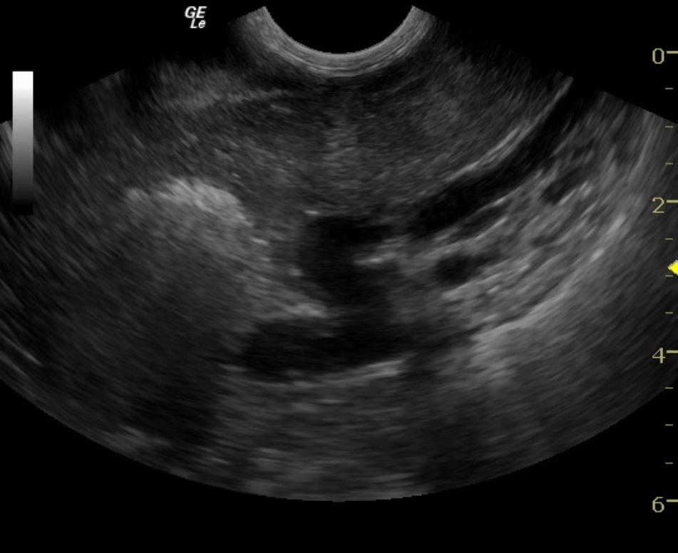
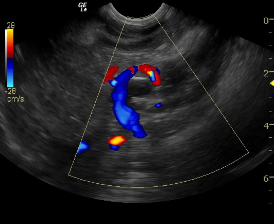
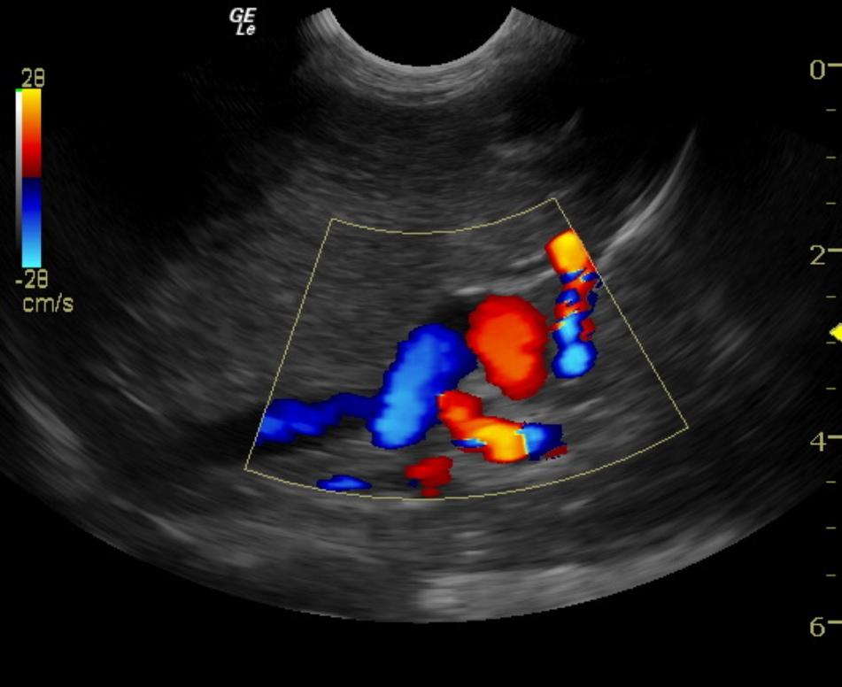
Image Interpretation
The liver was mildly subnormal in size. The parenchyma was hypovascular. The portal hilus in this patient revealed an extrahepatic shunt that appeared to be deriving from the portal vein with tortuous contour and measured 1.0 cm in width with dorsal direction with entrance into the thorax adjacent to the vena cava and aorta. This is strongly suggestive for splenoazygos shunt as the vena caval volume appeared normal and entered into the diaphragmatic inlet as did the aorta. The vena cava to aortic ratio was 1:1. The vena cava measured 0.8 cm at the portal hilus and also at the diaphragmatic inlet. The aorta measured 0.9 cm at the hilus and diaphragmatic inlet. Therefore, the most logical shunt would be an extrahepatic portoazygos shunt, likely splenoazygos in origin. The shunt appears to derive from the portal vein prior to the pyloric outflow cranial to the right kidney and dorsally directed. Ameroid constrictor measurement should be for a shunt that measured approximately 1.0 cm.
Outcome
Surgery was not performed. ACTH was normal. The patient is growing now, but thin. Shunt is suspected, but owners are not pursuing.
Video
Patient Information
Age : 2 Years
Gender : Female, Intact
Species : Canine
Status : Complete
Blood Chemistry
- Alkaline Phosphatase (SAP), High
- Azotemia
Clinical Signs
- Anorexia
- Renal Failure
- Vomiting
Images