A 2-year-old NM DSH was presented for evaluation of renomegaly on survey radiographs. Urinalysis and CBC were both normal. Mild azotemia was evident on serum biochemistry.
A 2-year-old NM DSH was presented for evaluation of renomegaly on survey radiographs. Urinalysis and CBC were both normal. Mild azotemia was evident on serum biochemistry.
Lymphoma, hydronephrosis, polycystic kidney disease, granulomatous disease, nephritis, pyelonephritis
FNA of the left kidney were performed without complication.
Strongly suggestive for bilateral renal lymphoma pattern. Dry form FIP is also possible, yet less likely.
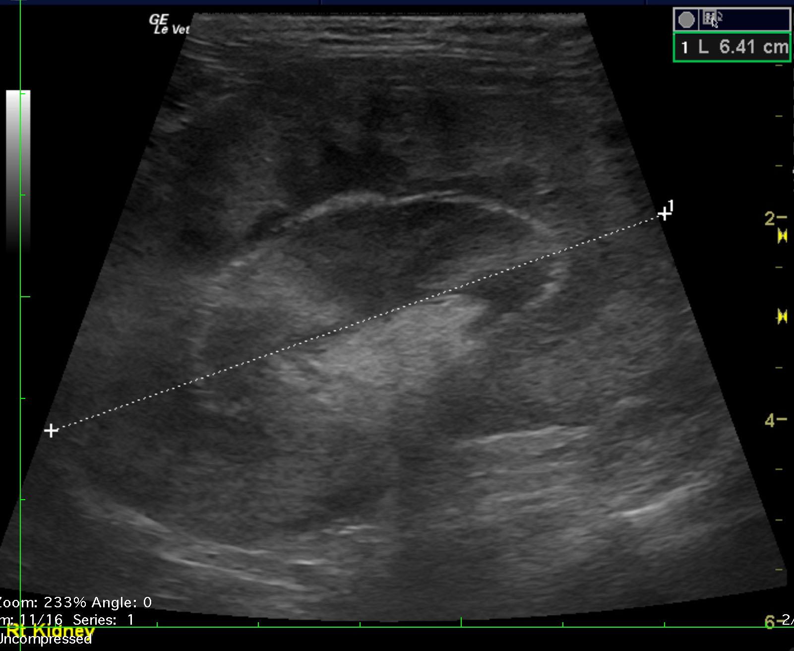
Hyperechoic medullary rim sign was noted along with ill-defined pelvic fat. Heterogenous, mixed nodular cortical changes were noted. This is strongly suspicious for renal lymphoma or dry form FIP. The right kidney was severely enlarged and measured 6.4 cm. The left kidney measured 7.64 cm with subcapsular halo with similar disruptive changes to the right kidney and more exaggerated nodular expansion.
None