A 6-year-old NM Dachshund with a history prednisone therapy for inability to walk was presented for evaluation of lethargy and anorexia. Abnormalities on CBC and serum biochemistry were anemia, band neutrophilia, azotemia, hyperphosphatemia, hypoproteinemia, elevated amylase, and prolonged PT and PTT.
A 6-year-old NM Dachshund with a history prednisone therapy for inability to walk was presented for evaluation of lethargy and anorexia. Abnormalities on CBC and serum biochemistry were anemia, band neutrophilia, azotemia, hyperphosphatemia, hypoproteinemia, elevated amylase, and prolonged PT and PTT.
GIT – ulceration, neoplasia, foreign body
Renal – acute kidney injury, chronic kidney disease, pyelonephritis, neoplasia
Peritonitis
Hemangiosarcoma – spleen, liver
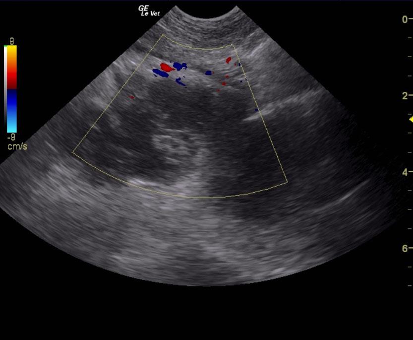
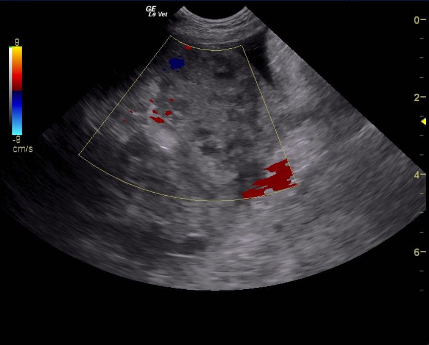
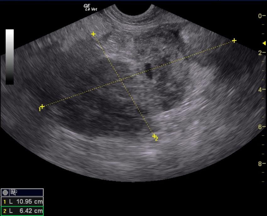
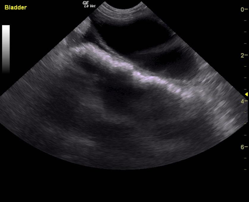
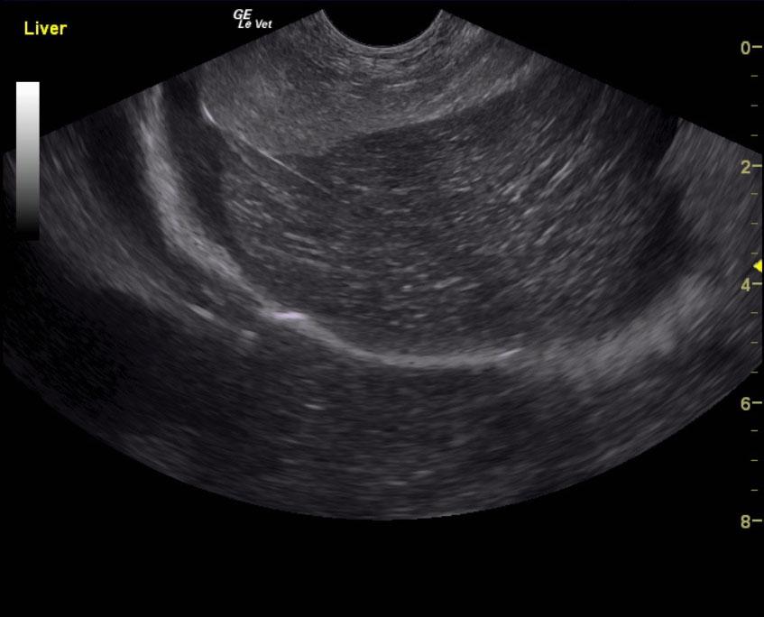
Left kidney mass and retroperitoneal extension, suspect hemangiosarcoma or similar neoplasia. Free fluid, consistent with hemoabdomen. A surgical consult can also be considered; however, blood transfusion is essential in this case. No overt evidence of metastatic disease was noted. It is possible that this may be represent a blood clot; however, it appears to be too coarse, too organized and deriving from the caudal pole of the left kidney.
Echocardiogram and chest radiographs are recommended. The coagulopathy in this case could be primary or secondary owing to consumptive disease from subacute bleeding over the last number of days from the renal mass. The mass will merit surgical resection regardless. Blood transfusion and plasma transfusions will be necessary in this patient to stabilize bleeding.
The right kidney presented mild, degenerative changes and corticomedullary mineralization. The renal length measured 4.8 cm. The left retroperitoneal space in this patient revealed an 11+ cm mass that was deriving from the caudal pole of the left kidney. This appears to be significantly structural and not necessarily a blood clot. Free fluid, associated with hemorrhage was noted in small pockets throughout the abdomen. FNAs are recommended after blood transfusion.