An 8-year-old NM DSH was presented for evaluation of anorexia and vomiting. Abnormalities on serum biochemistry were severe azotemia, hyperphosphatemia, and elevated total protein of 92.
An 8-year-old NM DSH was presented for evaluation of anorexia and vomiting. Abnormalities on serum biochemistry were severe azotemia, hyperphosphatemia, and elevated total protein of 92.
Acute kidney injury, chronic kidney disease, obstructive uropathy, pyelonephritis, FIP, renal lymphoma
Fluid from the kidney was obtained and submitted for culture. After 72 hours there was no growth.
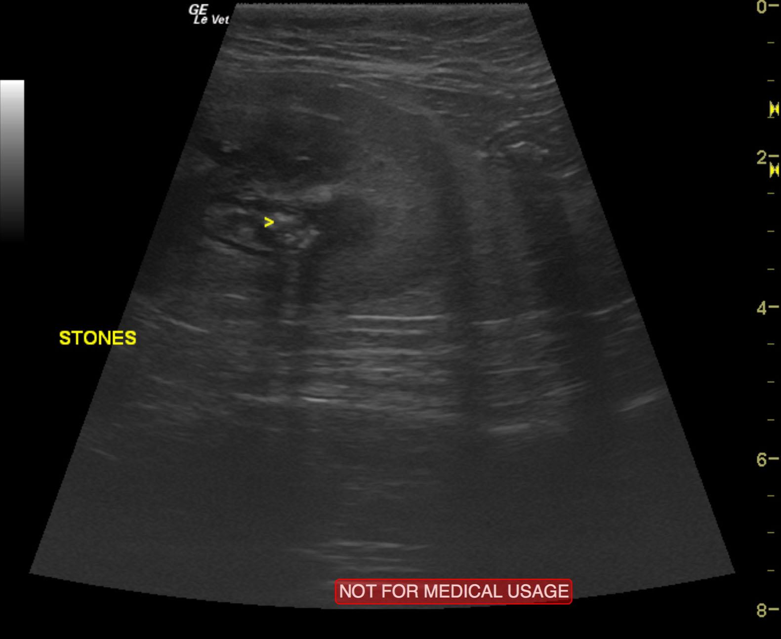
Right kidney pyelonephritis pattern with multiple calculi and pericapsular inflammatory pattern.
Chronic nephritis in the left kidney.
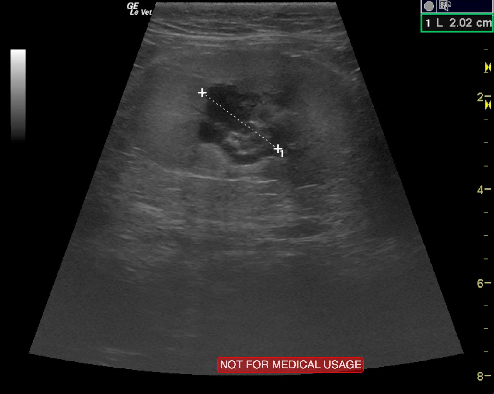
The right kidney was moderately enlarged with pericapsular inflammatory pattern. Pyelectasia was noted and measured 2.02 cm with multiple calculi within the right renal pelvis. The ureter was dilated approximately 1.0 cm beyond the kidney. No overt stone was noted in the ureter at this point. However, echogenic debris was noted. This would be suggestive of likely infection. The left kidney was small and measured 1.93 cm with pericapsular inflammatory pattern. This is consistent with low-grade chronic active nephritis.
None