06_00345 Marley T Bilateral pyelectasia

History

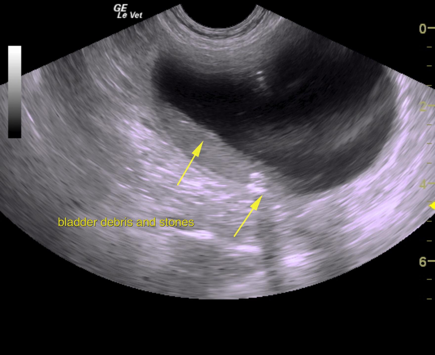
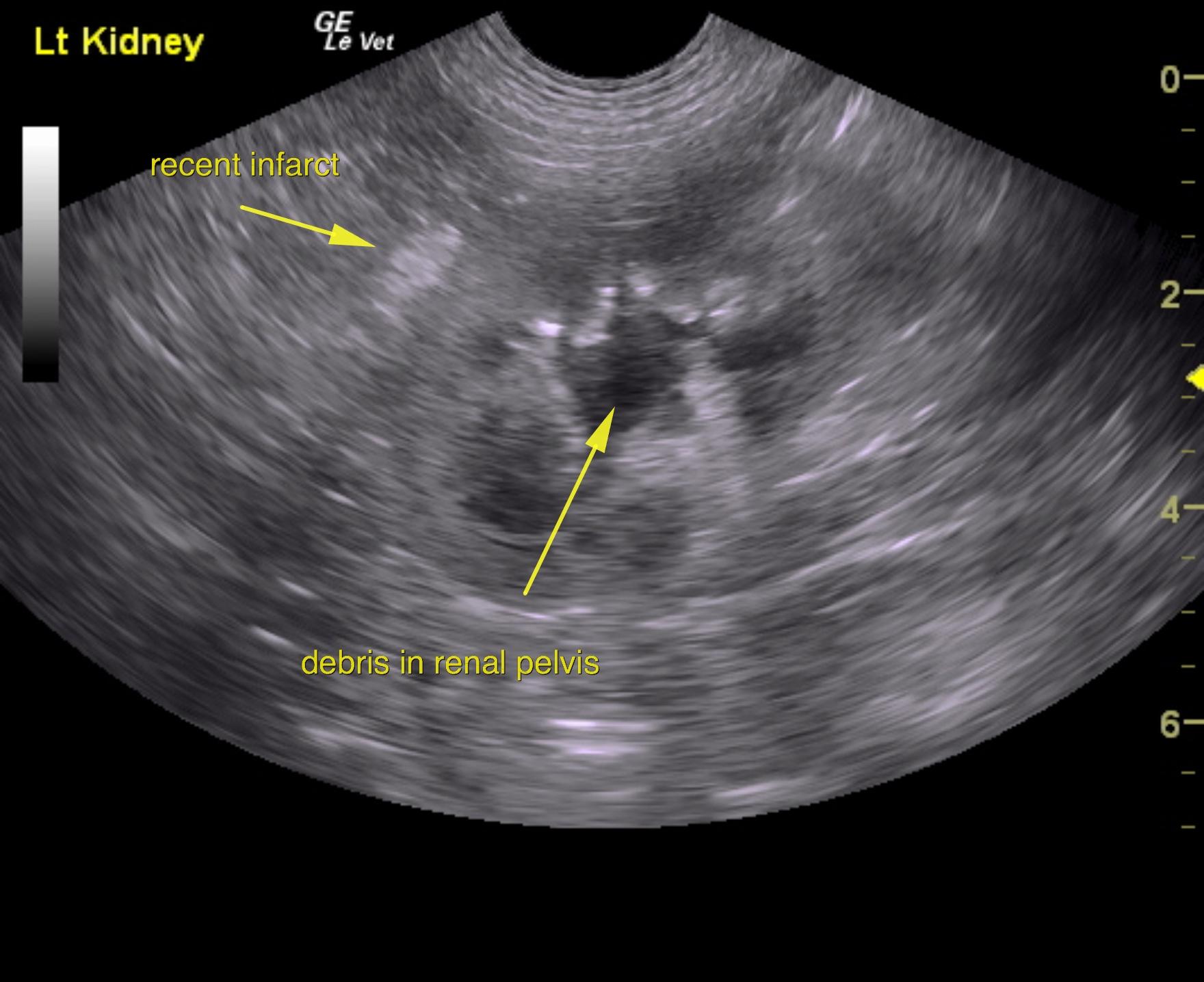
A 5-years-old male neutered Shih Tzu was presented for evaluation of urethral obstruction that had been managed with catheterization. Urinalysis showed SG of 1.008 and proteinuria.  Abnormalities on CBC and serum biochemistry were neutrophilia and monocytosis, elevated phosphorus, and azotemia; the latter had improved after being catheterized.


Clinical Differential Diagnosis

Urethral obstruction, chronic kidney disease, renoliths, pyelonephritis

DX

Bilateral pyelectasia

Sampling

None

Sonographic Differential Diagnosis

Bilateral renal pyelectasia. Occult infarct in the left kidney. Multiple renal calculi.
Small bladder calculi and debris.

Image Interpretation

The urinary bladder presented a large amount of debris. Urinary catheter was in place and appeared to be in proper position. Small calculi were noted in the bladder and were non-obstructive at this time. A slight amount of free fluid was noted cranial to the urinary bladder.
The prostate was slightly enlarged at 2.0 cm with dilated pelvic urethra and heterogenous parenchymal changes.
The right kidney measured 5.3 cm. Color flow assessment of the kidneys appeared to be adequate.
The kidneys in this patient presented pyelectasia that measured 0.95 cm in the left. Pyelectasia revealed echogenic debris. This is suggestive for pyelonephritis or potential occult pyelonephritis with washout given the PU/PD. Pinpoint calculi were noted with irregular contour and focal hyperechoic periserosal fat and indentation of the cortex. This is suggestive for infarct.

UA Specific Gravity Range

1.008

Outcome

None

Video

Patient Information

Age : 5 Years
Gender : Male, Neutered
Species : Canine
Status : Complete

Blood Chemistry

  • Azotemia
  • Phosphorus, High

CBC

  • Monocytes, High
  • Neutrophils, High

Images

mar6mar3mar4mar2mar7mar8marl1

Skip to content