A 13-year-old SF mixed breed dog with a previous complainant of urinary incontinence was presented for evaluation of PU/PD. Physical examination was within normal limits. Abnormalities on serum biochemistry were elevated ALT activity, and mild azotemia (creatinine 1.6, BUN 29).
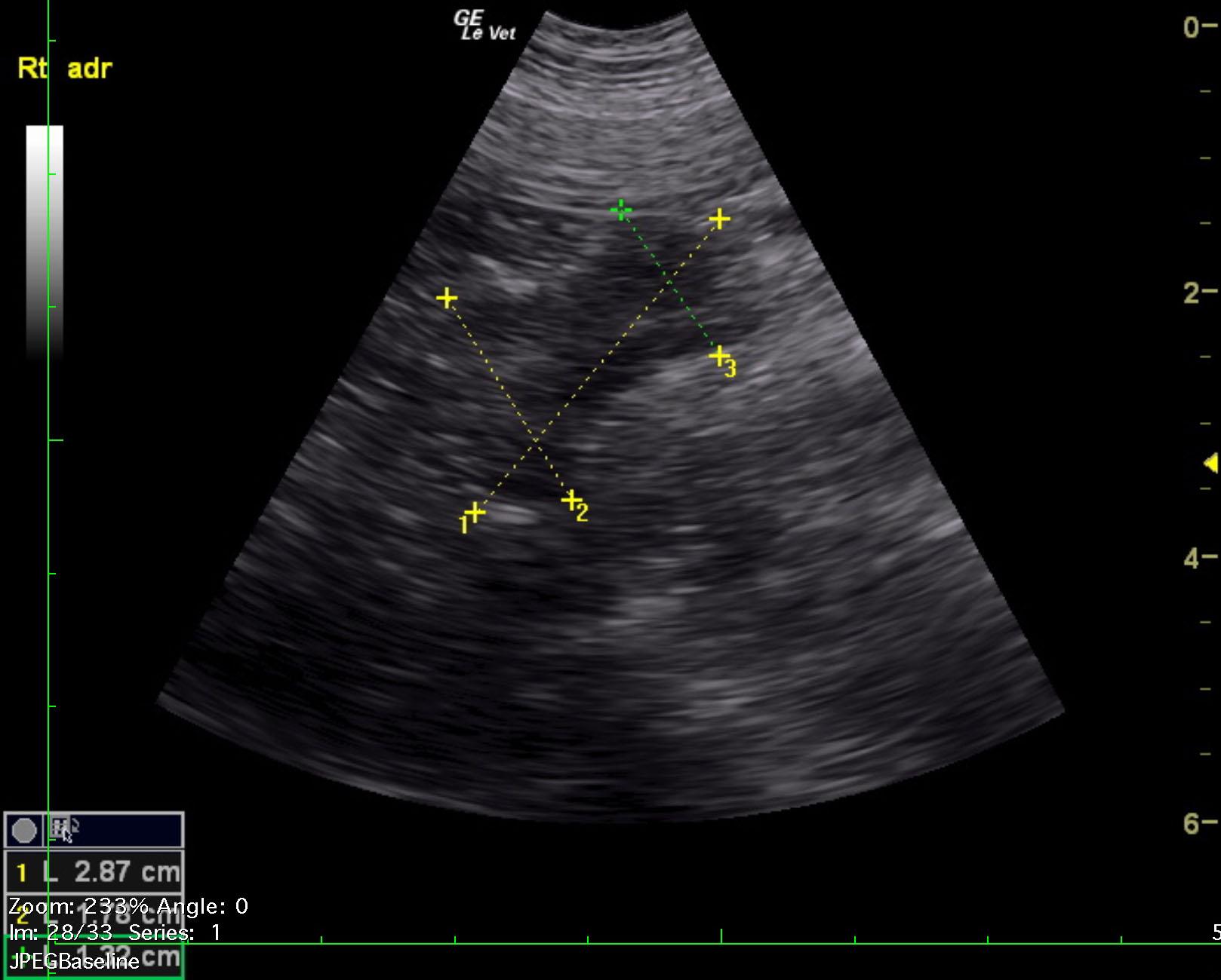
Right adrenal gland mass.
No evidence of caval invasion, likely phrenic vein invasion. This appears resectable. Pheochromocytoma, adenocarcinoma are possible.
Image Interpretation
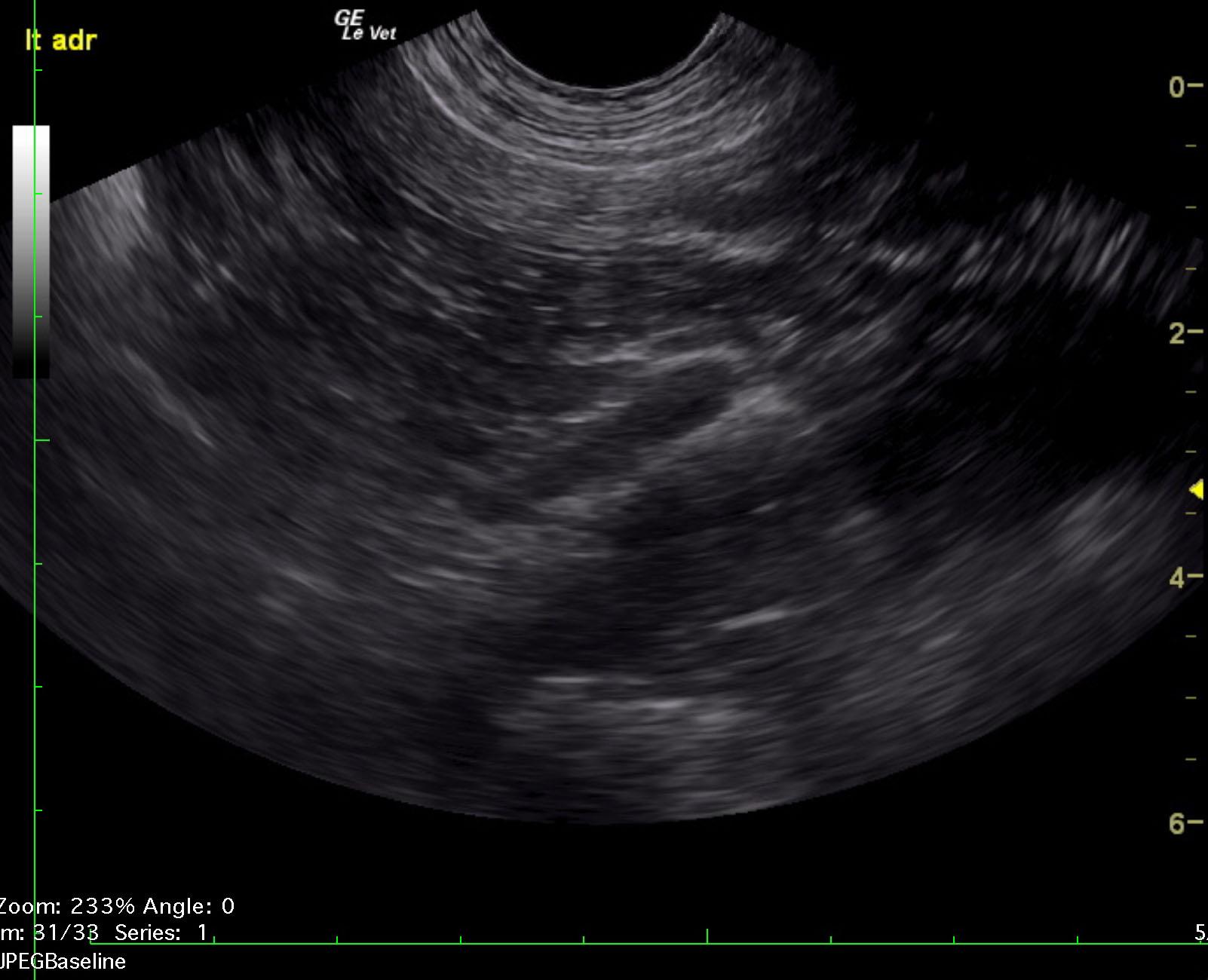
The left adrenal gland was uniform and measured 2.66 x 0.63 cm at the cranial pole and 0.59 cm at the caudal pole. The right adrenal gland was irregular, enlarged and hypoechoic measuring 2.87 x 1.78 cm at the cranial pole and 1.32 cm at the caudal pole. Irregular capsular expansion was noted. Vascularity around the right adrenal gland appeared to be relatively normal. However, the phrenic vein appeared to be slightly invaded or occupied by attached thrombus.