A 7-year-old NM DSH was presented for evaluation of PU/PD and weight loss. Survey radiographs showed small kidneys with mineralization and volume contracted heart. CBC and serum biochemistry showed anemia, hypoalbuminemia, hyperglobulinemia, azotemia, and hyperphosphatemia.
A 7-year-old NM DSH was presented for evaluation of PU/PD and weight loss. Survey radiographs showed small kidneys with mineralization and volume contracted heart. CBC and serum biochemistry showed anemia, hypoalbuminemia, hyperglobulinemia, azotemia, and hyperphosphatemia.
Adrenal mass in a 7 year old MN DSH cat
History
Comments
This mass is potentially resectable. However, due to the severe dystrophic changes of the left kidney, it is unclear if the patient could maintain any degree of appropriate renal function after removal of the right kidney. Therefore, the prognosis is guarded to poor long term.
Clinical Differential Diagnosis
Renal disease – chronic kidney disease, pyelonephritis, renoliths, lymphoma, granulomatous nephritis (fungal, bacterial, FIP).
DX
Adrenal mass.
Sampling
FNA of the adrenal gland was performed for further definition. Unfortunately, the results are not available.
Sonographic Differential Diagnosis
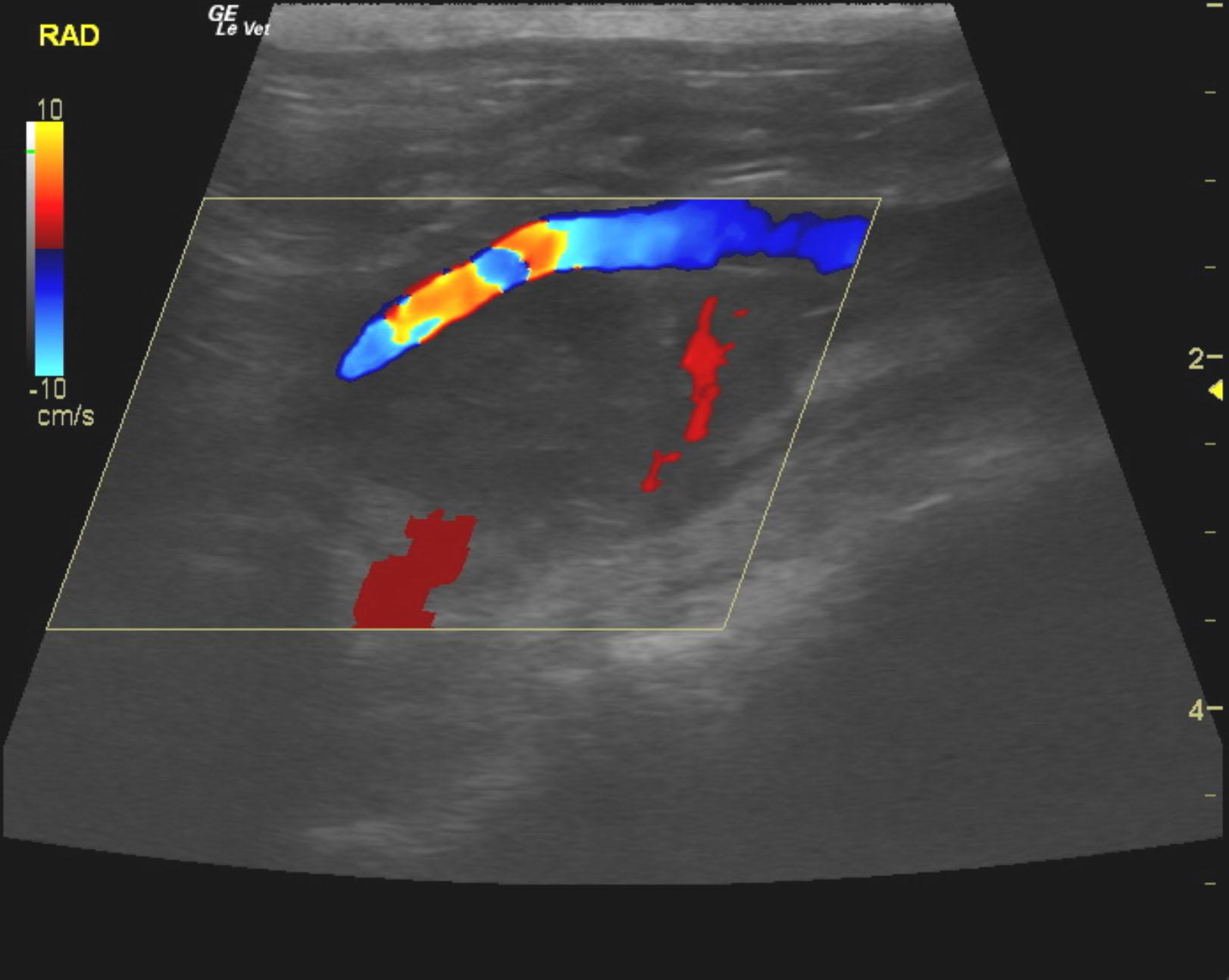
Mass in the area of the right adrenal gland encompassing or involving the right kidney. Adrenal invasion with involvement of the right kidney, or potential renal neoplasia with regional lymphadenopathy (obscuring the actual adrenal gland), is suspected.
Image Interpretation
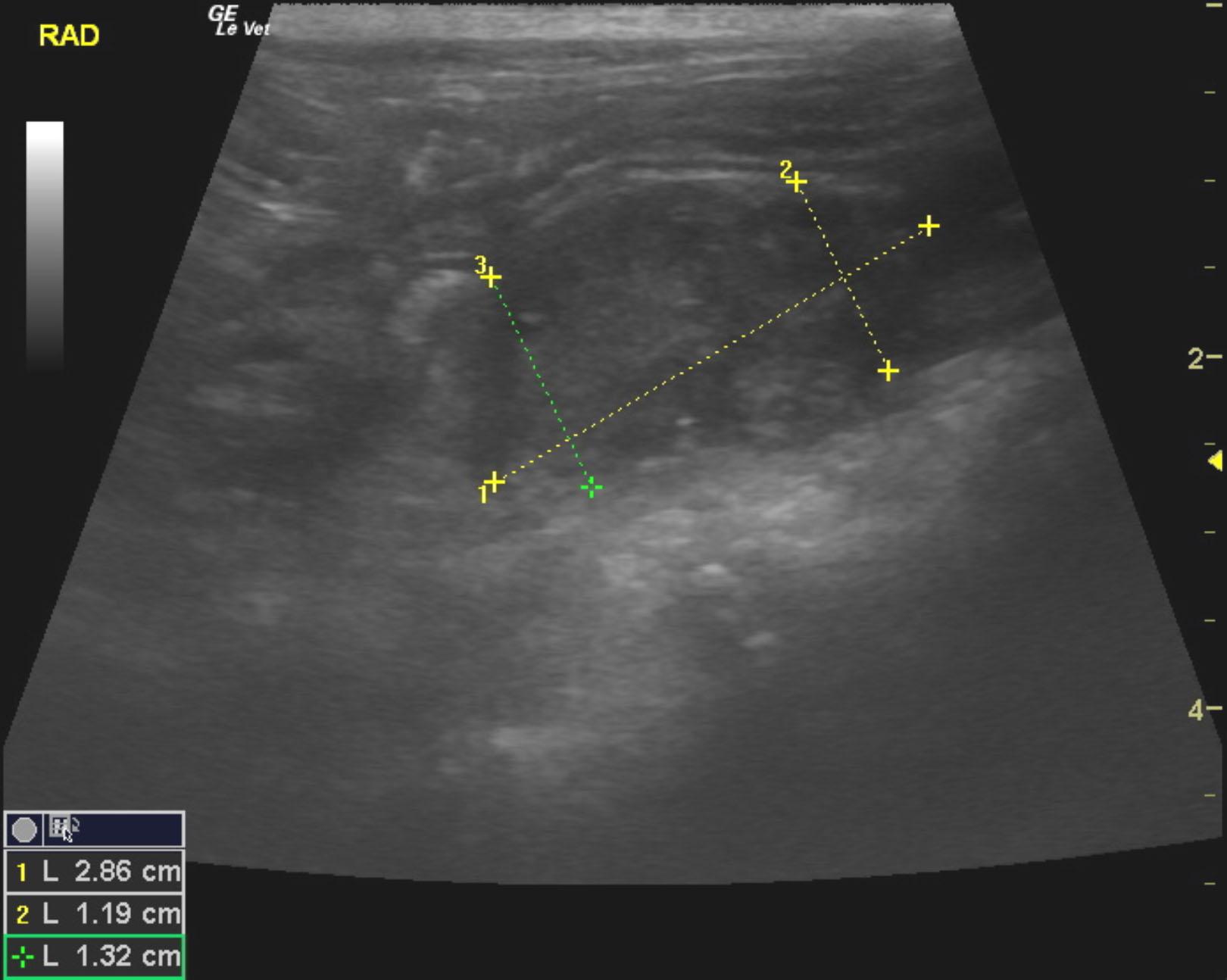
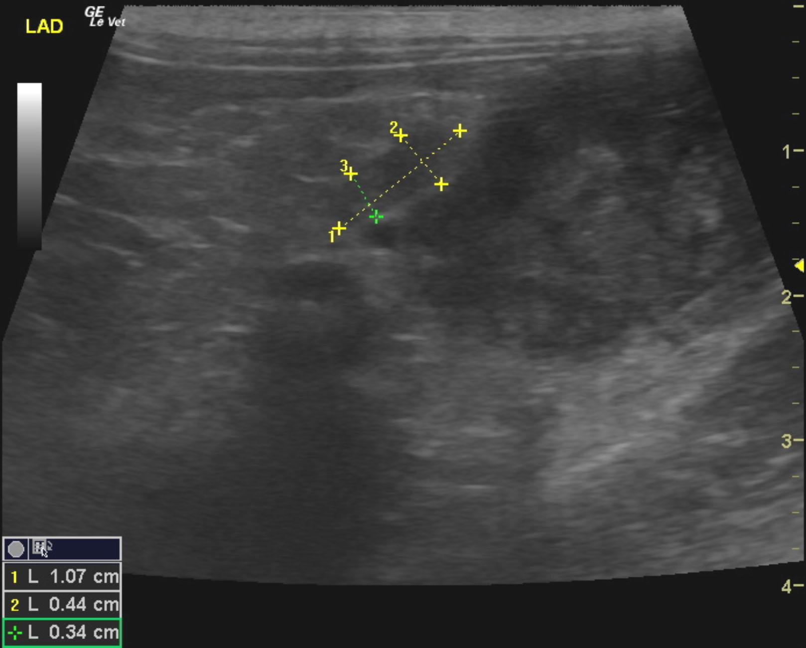
There was a mass noted in the region of the right adrenal gland adjacent to the right kidney that measured 2.86 x 1.32 cm. This mass appeared to be attached to the kidney. It was difficult to ascertain whether this mass was adrenal or lymph node in origin; an intimate relationship with the vena cava was noted, suggestive of an adrenal origin. Regardless, the mass meets neoplastic criteria. The left adrenal gland was uniform at 1.07 x 0.44 cm. There were severe dystrophic changes of the left kidney.
Outcome
None
Video
Patient Information
Patient Name : Mickey F
Age : 7 Years
Gender : Male, Neutered
Species : Feline
Liz Wuz Here : Yes
Status : Complete
Code : 07_00149
Blood Chemistry
- Albumin, Low
- Azotemia
- Globulin, High
- Phosphorus, High
CBC
- RBC, Low
Clinical Signs
- PU-PD
- Weight loss
Images