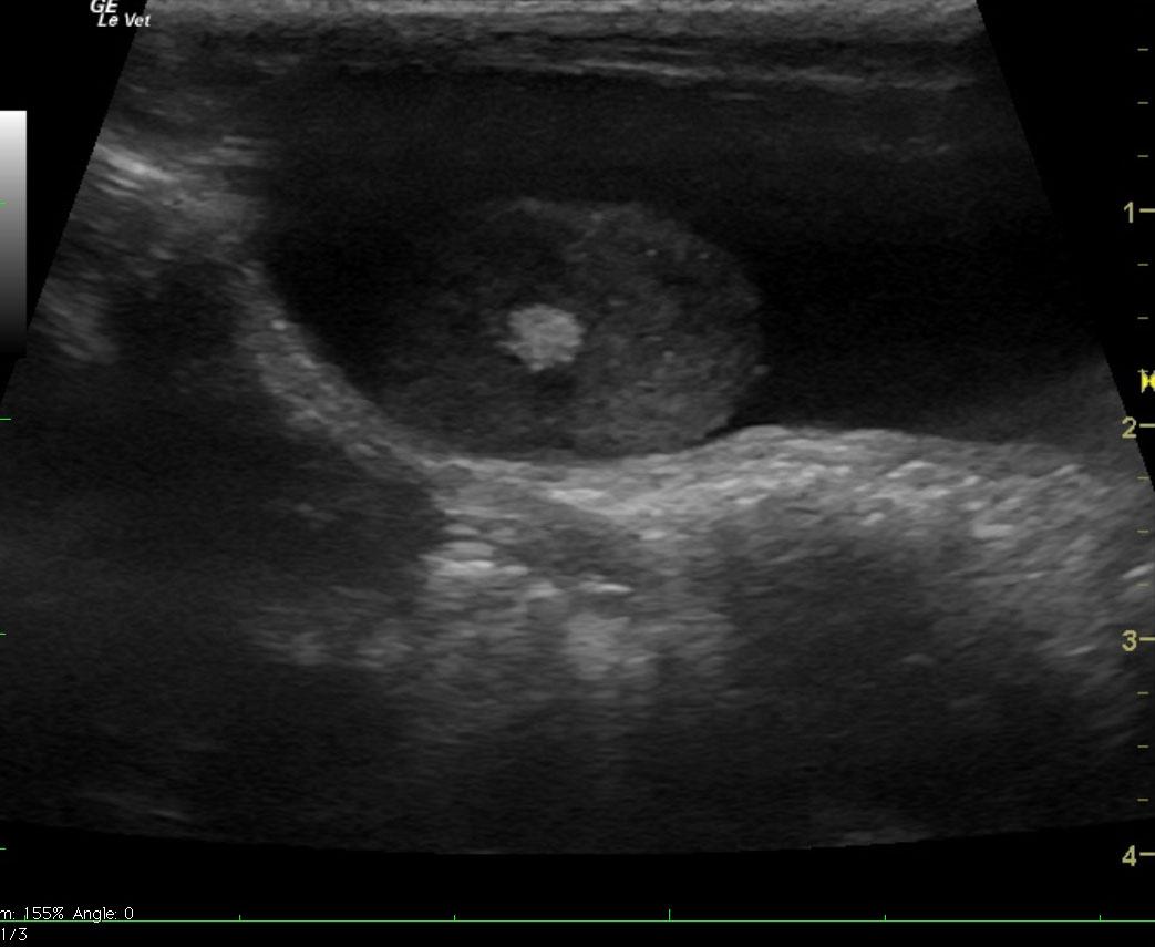
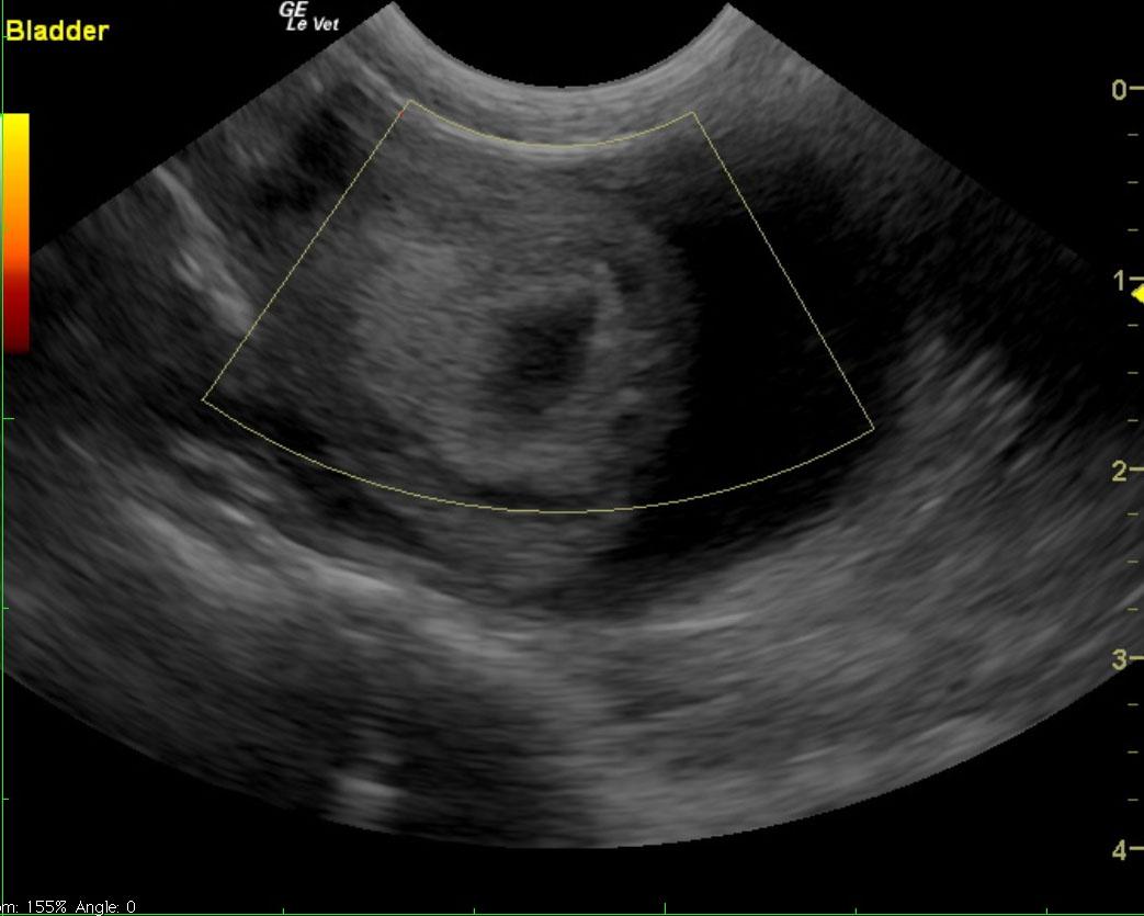
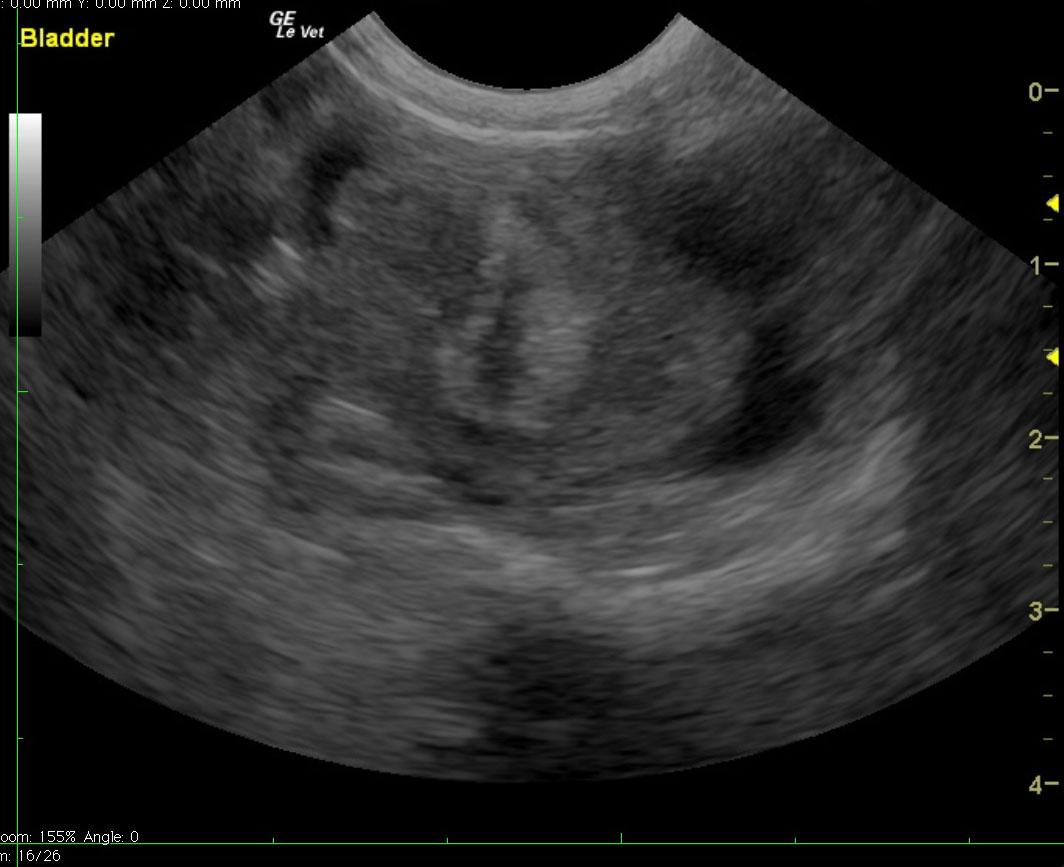
A 17-year-old NM Siamese was initially presented with PU/PD, increased appetite, and a bout of vomiting and diarrhea. Weight loss was noted on physical examination. The patient was later presented again with a hematuria. Bleeding from the penis, pale mucosa, and inappropriate mentation status (sitting sternal with head down) were present on physical examination. Urinalysis showed inappropriate SG (1.018), 1+ protein, and 3+ blood. Blood pressure, CBC, and coagulation panel were all within normal limits.
A 17-year-old NM Siamese was initially presented with PU/PD, increased appetite, and a bout of vomiting and diarrhea. Weight loss was noted on physical examination. The patient was later presented again with a hematuria. Bleeding from the penis, pale mucosa, and inappropriate mentation status (sitting sternal with head down) were present on physical examination. Urinalysis showed inappropriate SG (1.018), 1+ protein, and 3+ blood. Blood pressure, CBC, and coagulation panel were all within normal limits. Abnormalities on serum biochemistry included elevated ALT activity and amylase, and azotemia.