This 12 year old FS English Sheepdog has a hisotyr of nasal bleeding of 2 months duration
This 12 year old FS English Sheepdog has a hisotyr of nasal bleeding of 2 months duration
We use cookies to help you navigate efficiently and perform certain functions. You will find detailed information about all cookies under each consent category below.
The cookies that are categorized as "Necessary" are stored on your browser as they are essential for enabling the basic functionalities of the site. ...
Necessary cookies are required to enable the basic features of this site, such as providing secure log-in or adjusting your consent preferences. These cookies do not store any personally identifiable data.
Functional cookies help perform certain functionalities like sharing the content of the website on social media platforms, collecting feedback, and other third-party features.
Analytical cookies are used to understand how visitors interact with the website. These cookies help provide information on metrics such as the number of visitors, bounce rate, traffic source, etc.
Performance cookies are used to understand and analyze the key performance indexes of the website which helps in delivering a better user experience for the visitors.
Advertisement cookies are used to provide visitors with customized advertisements based on the pages you visited previously and to analyze the effectiveness of the ad campaigns.

This 12 year old FS English Sheepdog has a hisotyr of nasal bleeding of 2 months duration
This 12 year old FS English Sheepdog has a hisotyr of nasal bleeding of 2 months duration
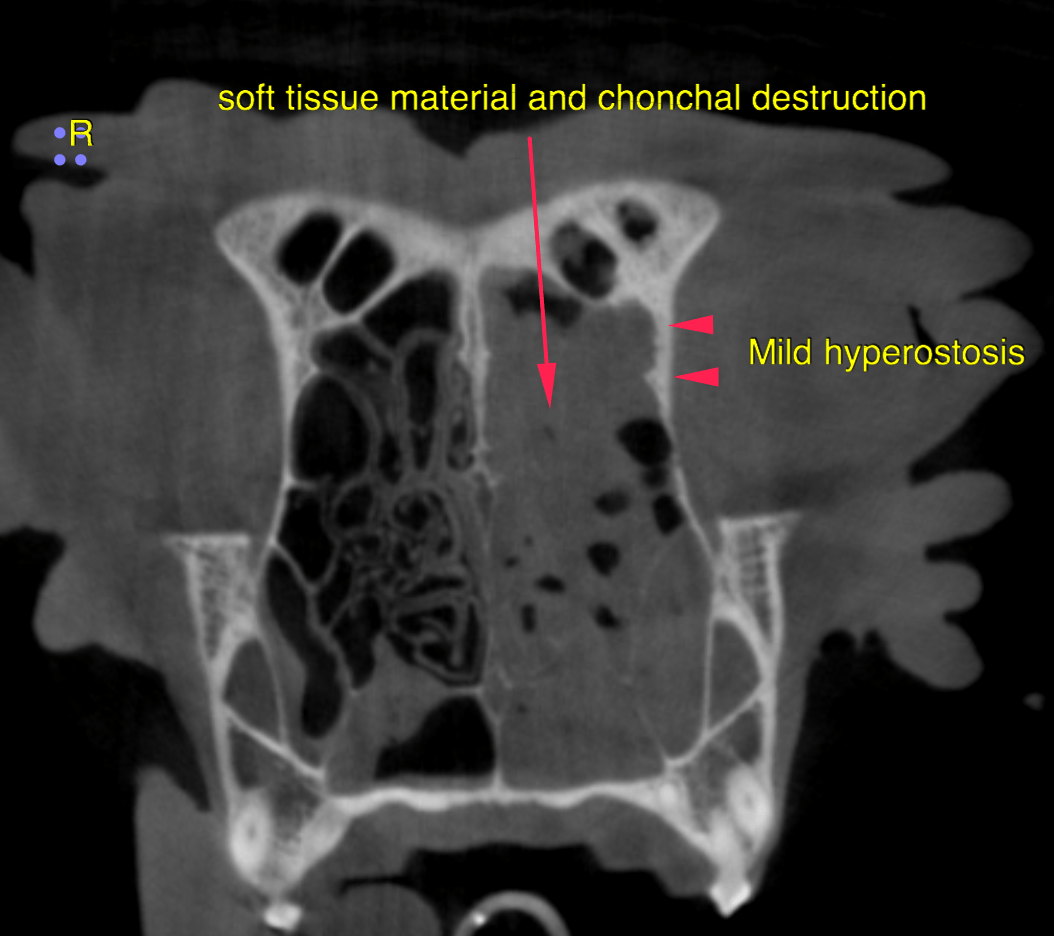
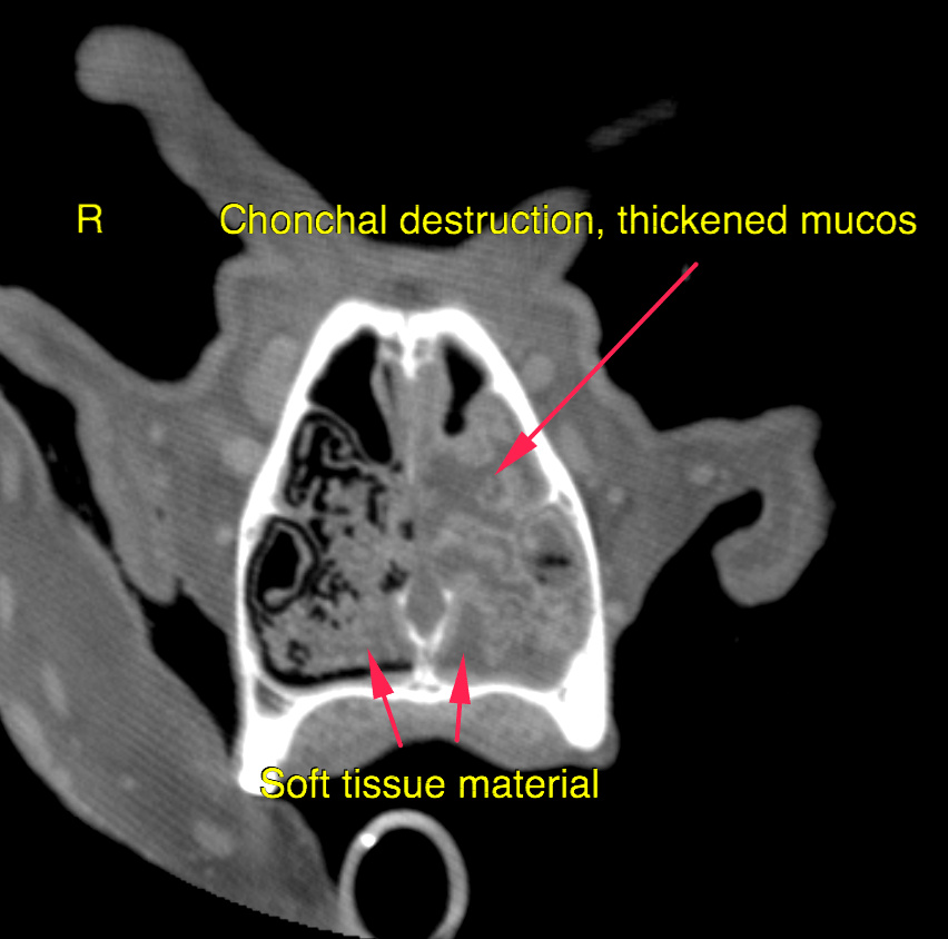
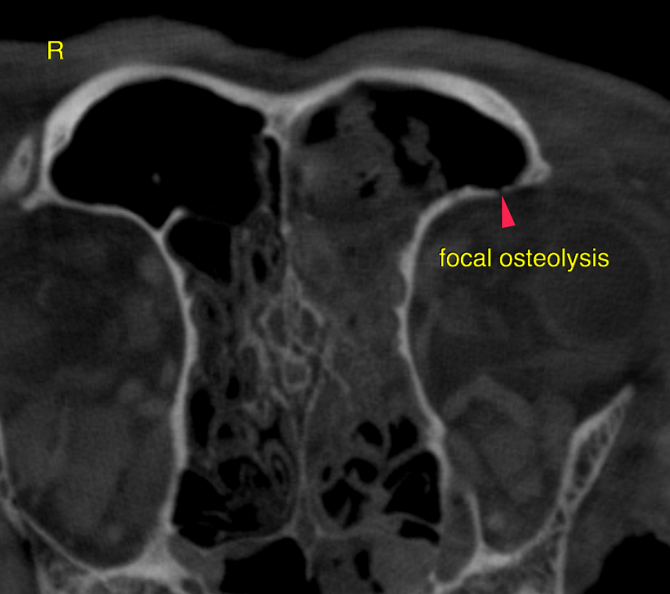
CT of the skull:
In the left nasal cavity there is a moderate amount of non-gravity dependent, noncontrast enhancing material present attached to the nasal turbinates. In the left frontalsinus and the nasopharynx a mild amount of gravity dependent material is present, demarcated by a horizontal fluid line. There is marked destruction of the conchal and turbinate structures in the left nasal cavity – consistent with an emerging “empty nose”. Mild turbinate destruction is noted within the right nasal cavity as well. The mucosal lining of the nasal cavity is moderately thickened with mildly increased contrast enhancement. The bony lining of the left nasal cavity presents multifocal mild hyperostosis and increased sclerosis. Small focal osteolytic areas are also noted.
As infectious agent Aspergillus is very likely.
Secondary reactive lymphadenitis left medial retropharyngeal lymph node is noted. Recommend rhinoscopy for further evaluation with sampling for culture and
histopathology (detection of fungi is more straightforward microscopically than with
culture in many cases) followed by antifungal therapy.