This 10 year old MN DSH cat has a history of controlled otitis media/externa AU, URI for the past 6 months. Some improvement with antibiotics but symptoms of wheezing, congestion and otitis return when stopped.
This 10 year old MN DSH cat has a history of controlled otitis media/externa AU, URI for the past 6 months. Some improvement with antibiotics but symptoms of wheezing, congestion and otitis return when stopped.
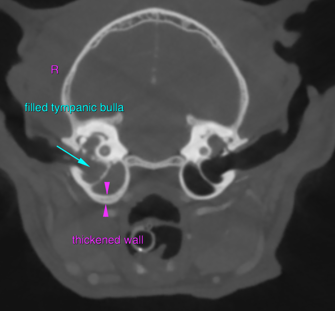
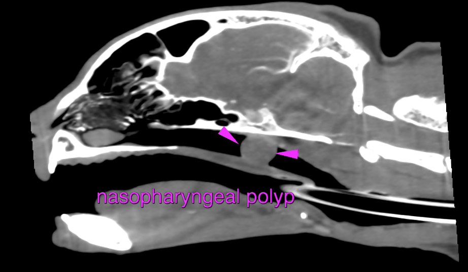
The right tympanic bulla presents a moderately thickened but smooth wall and contains uniform soft tissue attenuating and peripheral contrast enhancing material. In comparison to the contralateral side, the diameter of the osseous part of the auditory tube is moderately increased. A uniform soft tissue attenuating and peripheral contrast enhancing space occupying lesion is protruding from the right dorsolateral aspect of the nasopharynx – level with the opening of the auditory tube – into the lumen of the nasopharynx, occupying approximately 95% of its cross-sectional area.
Right sided chronic otitis media
Nasopharyngeal soft tissue mass, originating from the right tympanic bulla, causing upper airway obstruction
Secondary pressure induced mild atrophy of the osseous part of the right auditory tube
The findings are consistent with chronic inflammatory nasopharyngeal polyp originating from the right tympanic bulla.
Recommend – if not performed yet – rhinoscopy and removal of the nasopharyngeal polyp by traction technique. If clinical signs of clinical therapy are refractory after the polyp was removed, a right sided bulla osteotomy is recommended.