This 4 year old FS Schnauzer dog presented with congestion of 9 weeks duration. Other clinical signs include ataxia, running into walls, PU/PD, polyphagia and weight gain
This 4 year old FS Schnauzer dog presented with congestion of 9 weeks duration. Other clinical signs include ataxia, running into walls, PU/PD, polyphagia and weight gain
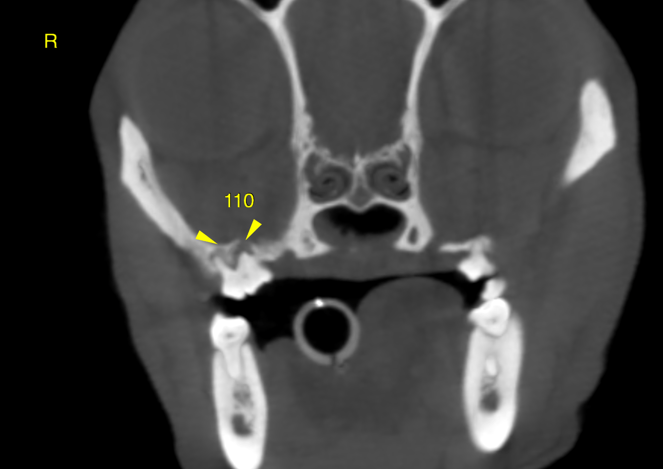
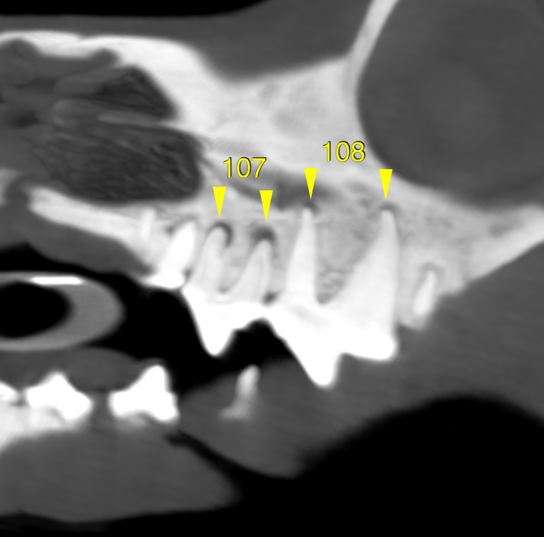
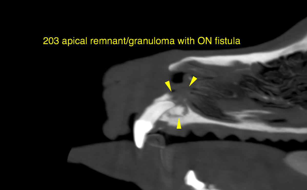
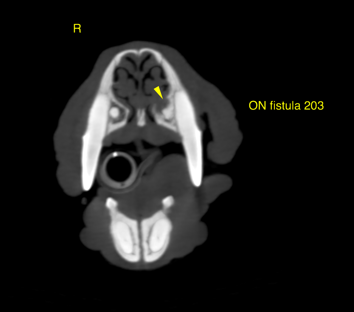
• Generalized periodontal disease with jaw bone atrophy • Apical tooth root infections 107, (108), 110 • Oronasal fistula with dentigerous cyst and small granuloma 203 • Mild unspecific non-destructive secretory rhinitis • Polypoid nasopharyngeal soft tissue mass Rightsided otitis media
CT of the head –
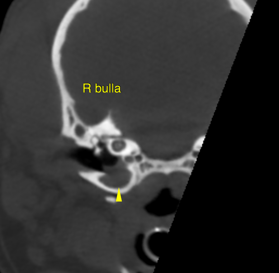
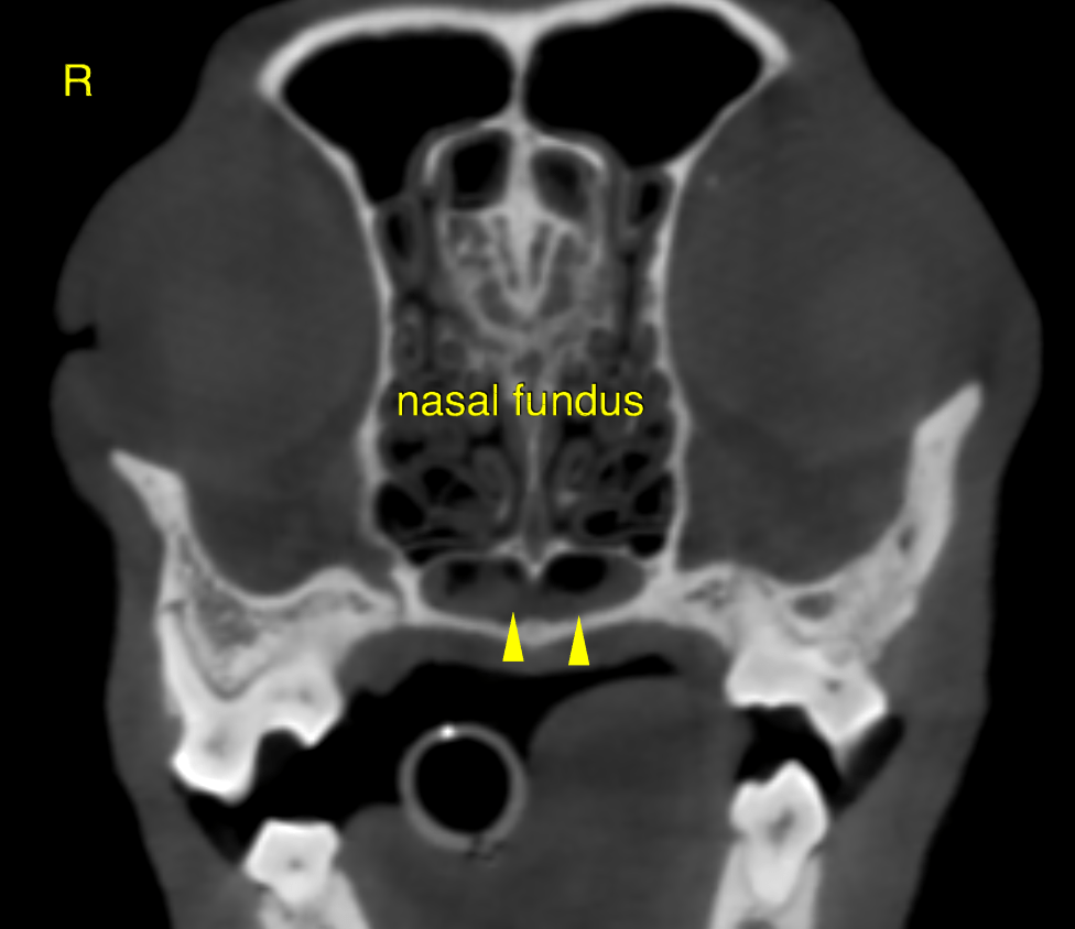
The computed tomography of the head reveals atrophy of the jaw bones and generalized periodontal disease with apical tooth root infections of the 107, 108 and 110. An oronasal fistula is seen at 203 with an impacted decidual tooth root remnant/dentigerous cyst and a small apical granuloma expanding into the nasal cavity. A mild amount of fluid nasal secretions is noted bilaterally within the ventral nasal meatus and nasal fundus. A polypoid well delineated soft tissue attenuating mass lesion of 3.5 mm diameter is emerging from the caudodorsal aspect of the nasopharynx in a midline position. The right tympanic bulla is partially filled with soft tissue attenuating material with a meniscal fluid gas interface indicating viscuous fluid.
Full dental and otoscopic workup is recommended. Excision of 203 including the dentigerous cyst and sealing oft he fistula is recommended. Consider extraction of 107 and 110 as well.
Differentials for the nasopharyngeal mass include inflammatory polyp as well as dermoid, or emerging neoplasia such as lymphosarcoma, melanoma, squamous cell carcinoma, other. Obtain biopsies for further definition. The lesion should be accessible via retrograde rhinos copy. Obtain samples for bacterial and fungal cultures as well. At this point the CT findings do not support desctructive forms of rhinitis such as fungal.