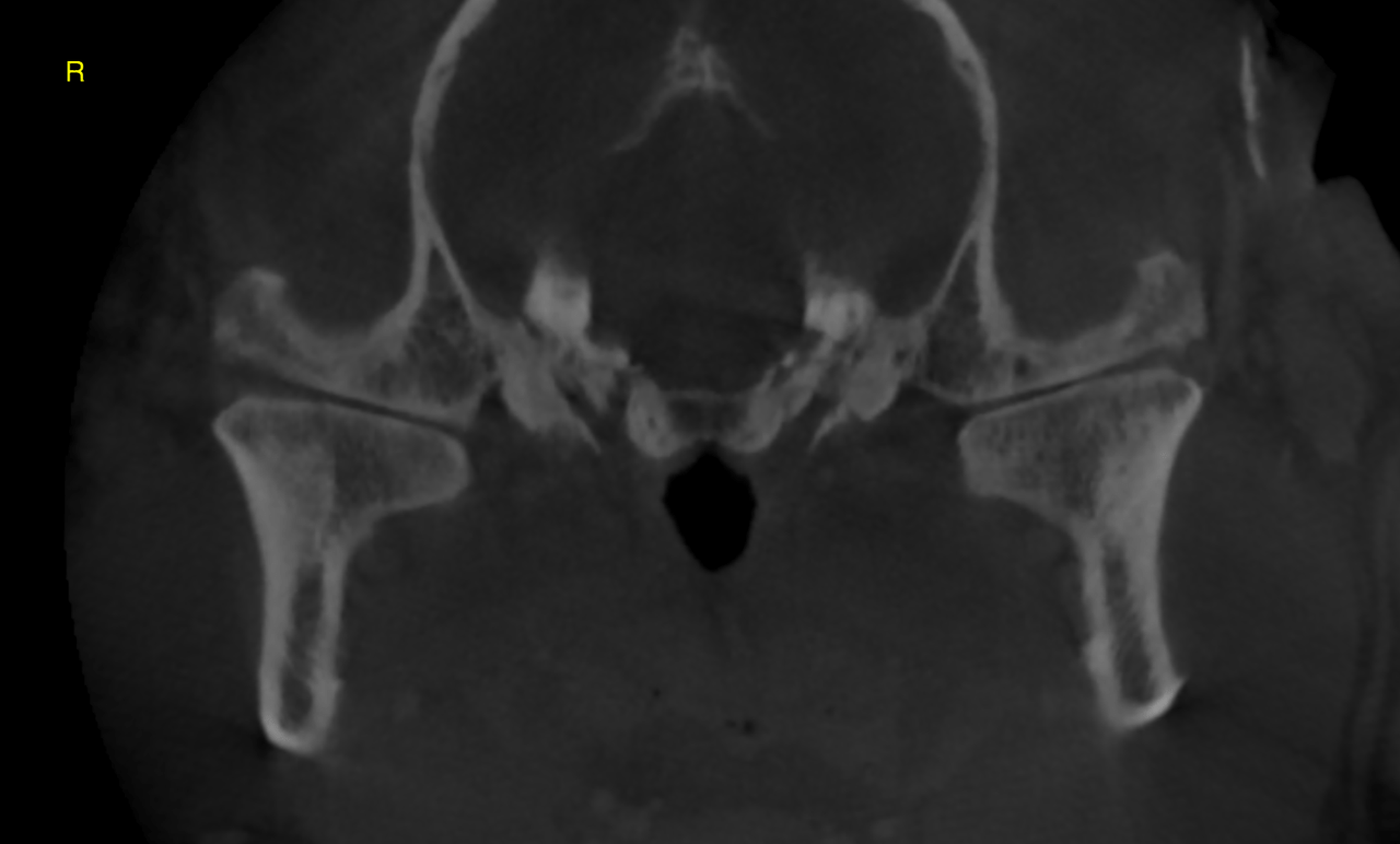
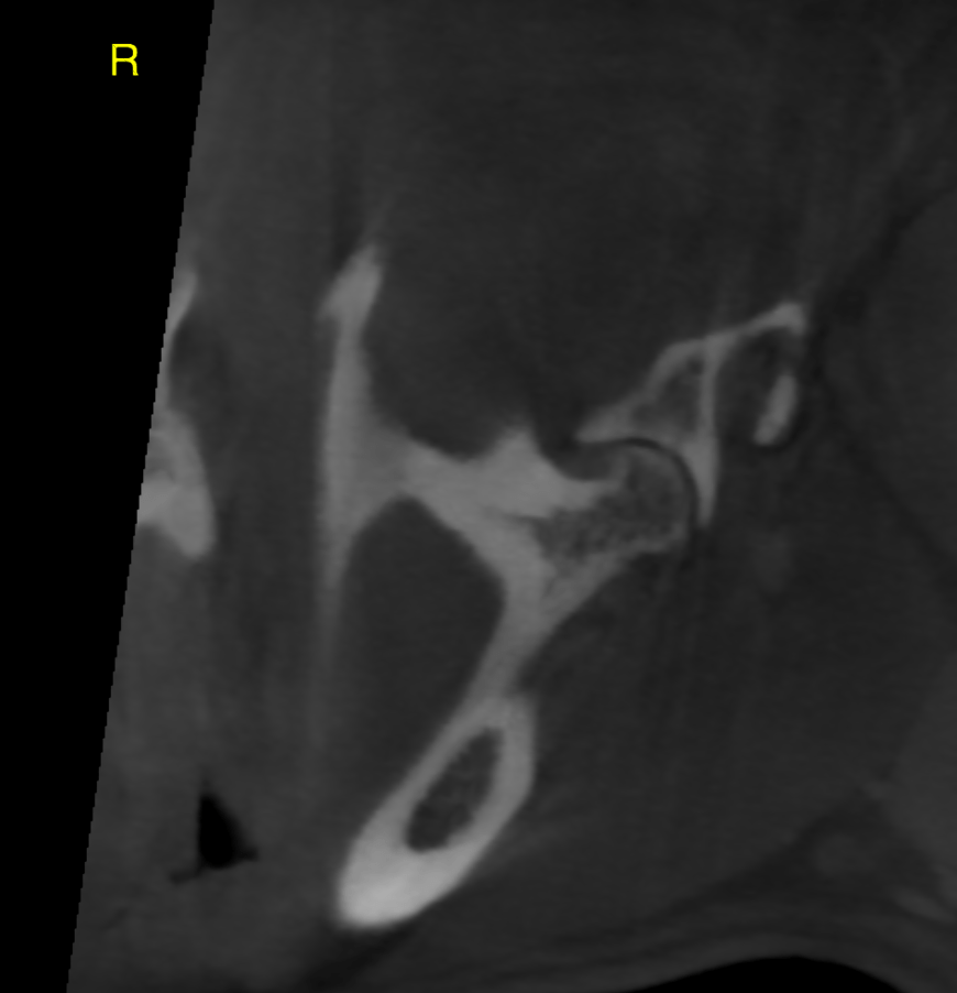
This 6 year old FS dog presented with a history of inability to open her mouth or eat starting 3 days prior. Appears to be acute onset of cx sign. No facial pain was noted or any CN abnormalities. Can only eat gruel at this time. No hx of maxillofacial trauma
Physical Exam: all vital signs wnl. able to manually open her mouth 7 mm (from apex of 103/203 to 304/404 tooth tips. no CN abnormalities. no facial pain. LNN palpate normal size and consistency. moderate periodontal disease. Obvious plaque, gingival hyperplasia, and gingival recession
CBC/Chem: globulin 5.1
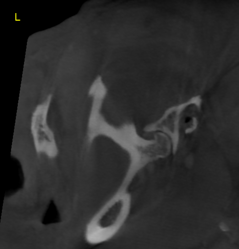
Reason for Ultrasound Exam: Has not been able to open mouth all the way.Want to R/O TMJ Disease vs Masticatory Muscle Myositis. 2M antibody test is pending.