CT – Pulmonary bulla rupture with spontaneous pneumothorax in a 6 year old MN Newfoundland
History
This 6 year old MN Newfoundland has a history of recurrent pneumothorax of one week duration, requiring 4 chest taps. Suspect pulmonary bulla.
This 6 year old MN Newfoundland has a history of recurrent pneumothorax of one week duration, requiring 4 chest taps. Suspect pulmonary bulla.
DX
The findings are consistent with history of a spontaneous pneumothorax after rupture of a pulmonary bulla.
Image Interpretation
CT of the thorax –
The lung lobes bilaterally are retracted from the thoracic wall and a mild to moderate amount of free gas is noted within the pleural cavities.
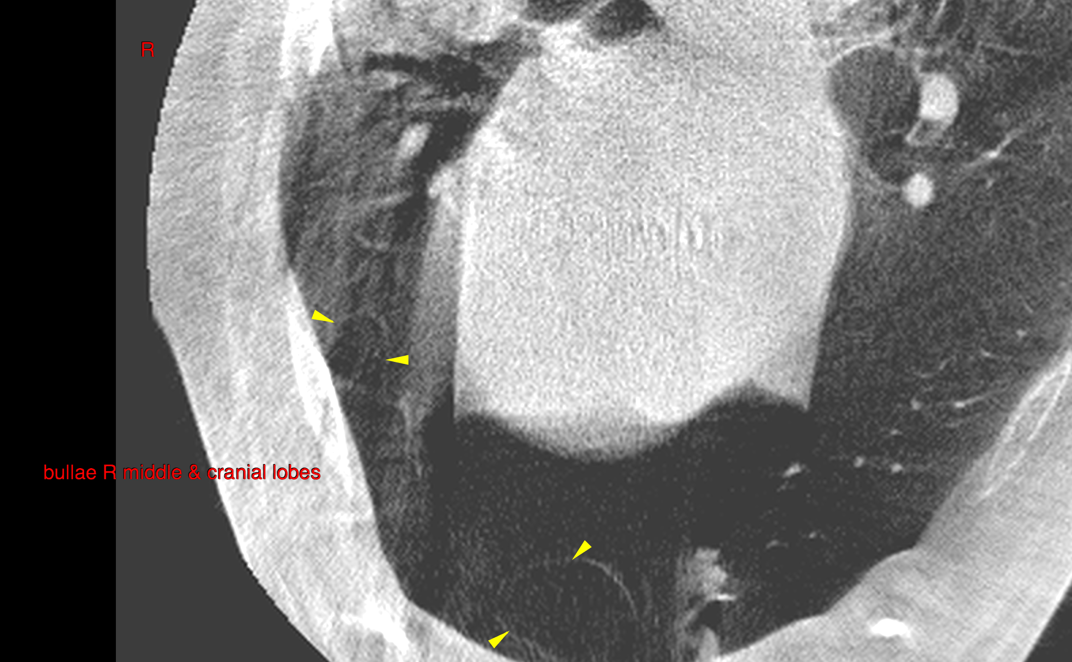
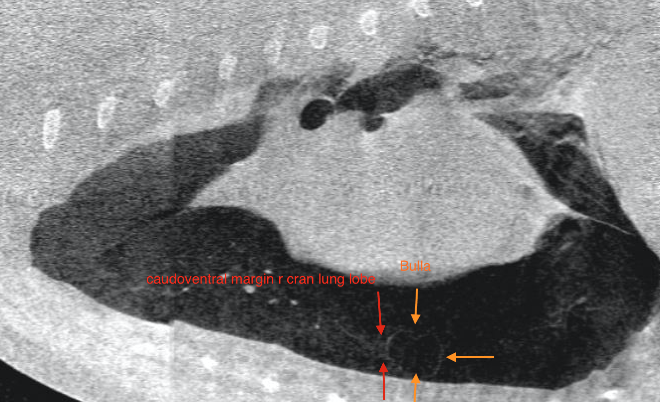
The ventrocaudal margin of the right cranial lung lobe presents a thin walled gas filled cavitary lesion of 28 mm diameter.
The ventral portion margin of the right middle lung lobe presents a thin walled gas filled cavitary lesion of 10 mm diameter.
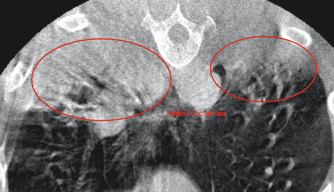
The dependent peripheral aspects of the lung parenchyma present ill-defined areas of soft tissue attenuation accompanied by a reduced volume. Airbronchograms are noted in these regions.
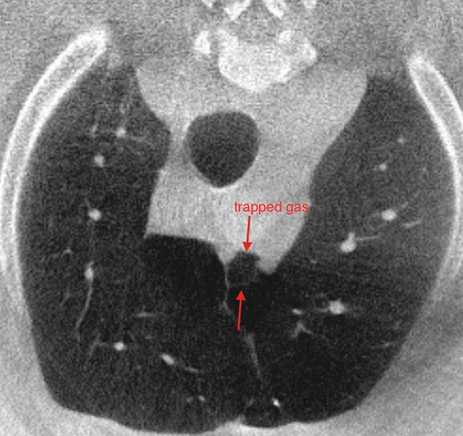
Small thin walled gas filled pleural folds are noted cranial to the heart base.
Outcome
Moderate bilateral pneumothorax
Cavitary lesions right cranial and right middle lung lobe
Compression atelectasis dependent areas of the lung, due to general anesthesia
Trapped gas in pleural and mediastinal folds
Incidental pleural scarring of the caudoventral mediastinal reflection
Lobectomy of the leaking lung lobe(s) has been performed already.