A 10-year-old neutered male Shih Tzu was presented for evaluation of intermittent stranguria and weight loss. On rectal palpation, there was some thickening of the prostate. Urinalysis and radiographs were both within normal limits, and blood work showed elevated ALP activity.
A 10-year-old neutered male Shih Tzu was presented for evaluation of intermittent stranguria and weight loss. On rectal palpation, there was some thickening of the prostate. Urinalysis and radiographs were both within normal limits, and blood work showed elevated ALP activity.
Bladder pathology – urolith, neoplasia, granulomatous cystitis. Prostatic disease – neoplasia, abscessation. Urethral disease – neoplasia, urethrolith. Increased ALP – vacuolar hepatopathy, nodular regeneration, neoplasia, granulomatous disease.
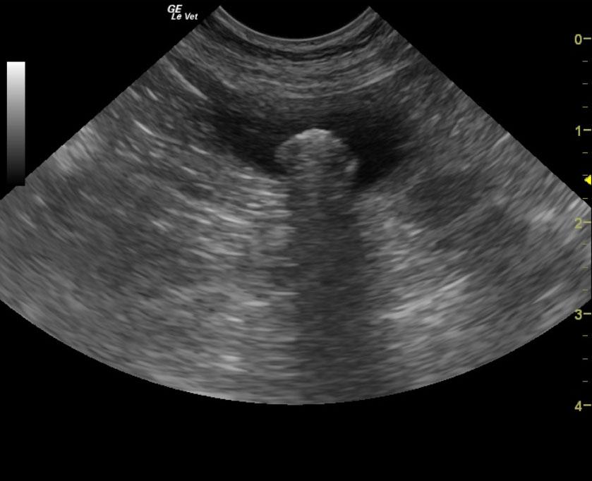
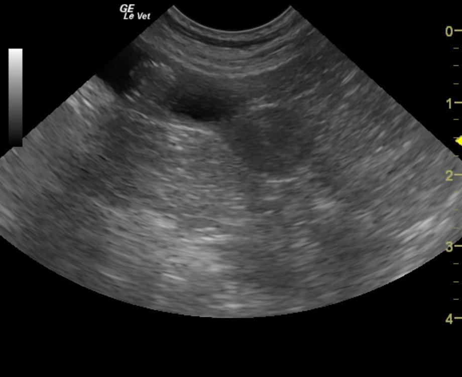
The urinary bladder was structurally unremarkable with anechoic urine and a normal bladder wall. However, a 1.0 cm shadowing cystic calculus was noted. See comments in the outcome section below for further discussion of the liver.
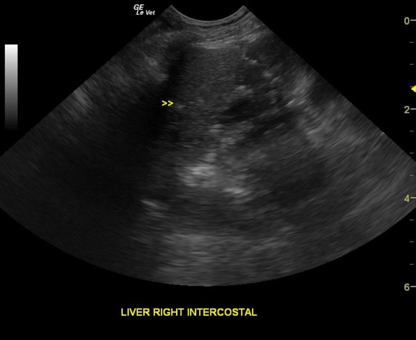
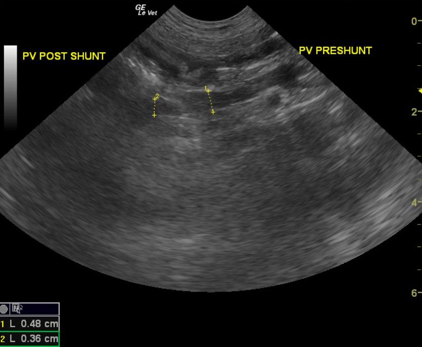
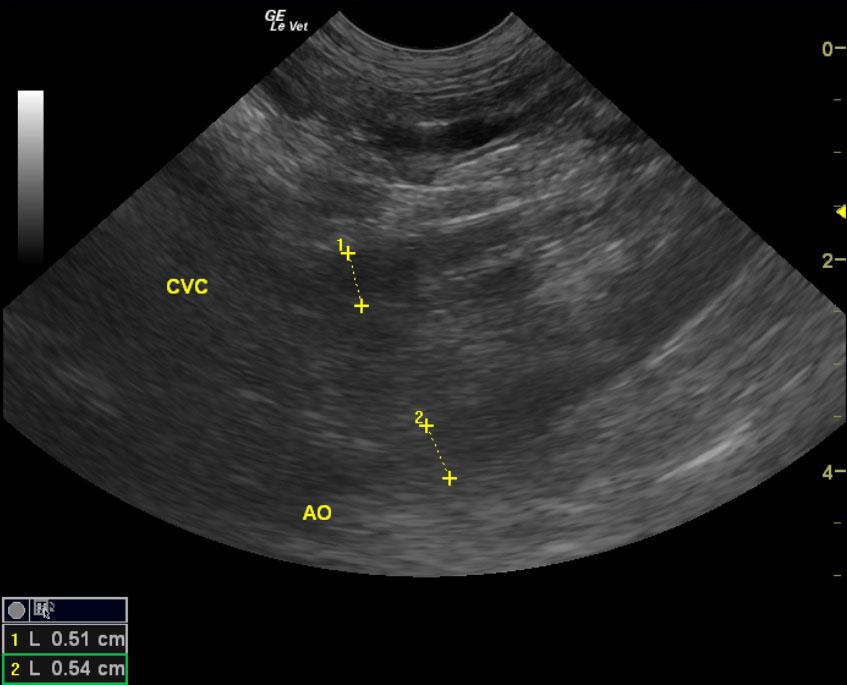
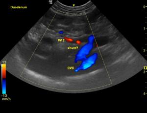
A repeat ultrasound was done a few days later. Further review of the liver on an empty stomach with less artifact in the region of the portal hilus revealed a 0.9 cm splenoazygos shunt and microhepatica with some remodeling of the liver. Pre shunt the portal vein measured 0.48 cm, post shunt 0.31 cm. The vena cava measured 0.51 cm, aorta 0.54 cm. The shunt was investigated again after finding that the cystotomy revealed an ammonium biurate calculus. The region was not overtly visible during the prior sonogram owing to artifact provided by the transverse and ascending colon and stomach.