The patient is a 6 year old FS PitBull dog who is non-weight-bearing on the right hind leg
The patient is a 6 year old FS PitBull dog who is non-weight-bearing on the right hind leg
The patient is a 6 year old FS PitBull dog who is non-weight-bearing on the right hind leg
The patient is a 6 year old FS PitBull dog who is non-weight-bearing on the right hind leg
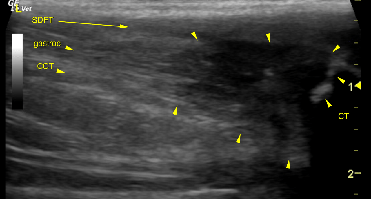
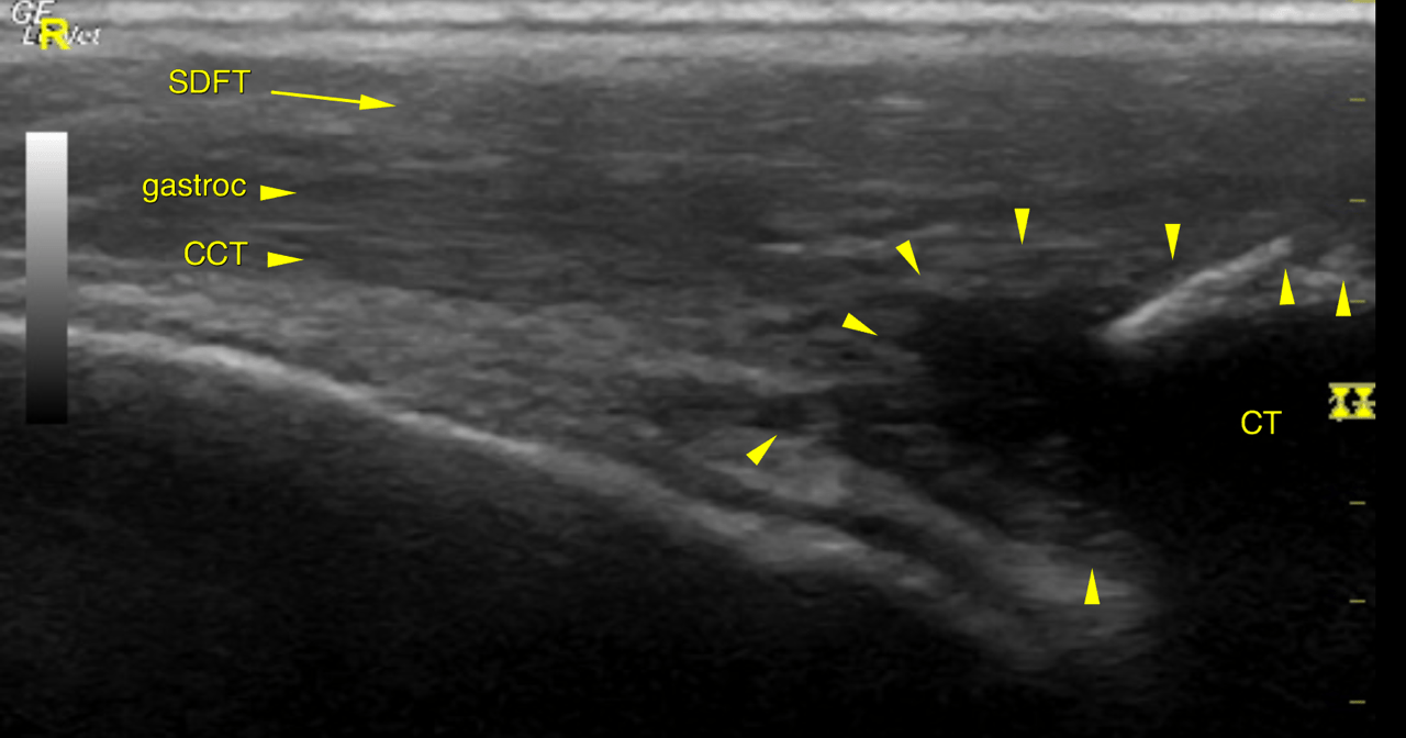
Ultrasound of the left and right calcanea tendons – Left:
Marked localized swelling with partial fibre disruption, decreased echogenicity and
increased heterogeneity is noted for the gastrocnemius and common calcaneal tendons
next to their calcaneal insertion. The affected region extends approximately 2 cm
proximal from the calcaneal tubercle and comprises hypo- to anechoic regions next to
echogenic foci.
The bone surface of the calcaneal tubercle presents marked irregularity and modeling
with concave defects and multiple protruding new bone formations.
The calcaneal bursa reveals mild enlargement and effusion. A minor amount of
anechoic fluid is seen between the tendinous components on the calcaneal tendon
localized to the area of fibre disruption.
The superficial digital flexor tendon reveals mild swelling and loss of echotexture
localized to the calcaneal tubercle.
Right: Moderate localized swelling with partial fibre disruption and markedly decreased
echogenicity is noted for the common calcaneal tendon and part of the gastrocnemius
next to their calcaneal insertion. The affected region extends approximately 1 cm
proximal from the calcaneal tubercle and comprises hypo- to anechoic regions.
The bone surface of the calcaneal tubercle presents moderate irregularity and
modeling.
A minor amount of anechoic fluid is seen between the tendinous components on the
calcaneal tendon localized to the area of fibre disruption.
The superficial digital flexor tendon reveals mild swelling and loss of echotexture
localized to the calcaneal tubercle.
The findings are more advanced on the left side; acute, subacute and chronic partial
rupture stages are present simultaneously. On the left side the entire cross section of
both the common calcaneal tendon and gastrocnemius tendon are affected. On the right
side the entire cross section of the common calcaneal tendon but only part of the
gastrocnemius tendon are affected. The ultrasonographic changes mainly suggest
subacute and acute rupture stages here.
Chronic bursitis (left) and tendinitis of the superficial digital flexor tendon (left &
right) are noted as additional findings.
Generalized degeneration with repetitive microruptures – such as in Doberman- is is
most likely here since the disease is bilaterally present and occurs at a typical age.
Rule out hypothyroidism, Diabetes and hyperadrenocorticism as underlying disease.
Lameness, toe tipping stance & gait typically develop at the point of biomechanical
failure.
At this relatively advanced stage response to conservative management is unlikely.
Successful healing requires temporary fixation.
Ultrasonographic monitoring of the tendon healing should be considered. Restoration
of longitudinal fibres should be ensured before the fixation is discontinued.