This 9 year old FS Border Collie dog presented with Chronic intermittent left hind limb lameness; varies from off weights to NWB. Does agility, has had multiple sessions of laser therapy, recently anaplasmosis positive, treated with Doxy. Recently started Adequan.
This 9 year old FS Border Collie dog presented with Chronic intermittent left hind limb lameness; varies from off weights to NWB. Does agility, has had multiple sessions of laser therapy, recently anaplasmosis positive, treated with Doxy. Recently started Adequan.
PE: Left proximal satorius ropey on palpation, pain on palpation. Left stifle effusion noted 1/13, not on 2/2. Iliopsoas body and insertion pain. Patient extremely limited to extend stifle or hip, or allow soft tissue stretches. She also has compensatory back pain and spasms that were not present prior to LR lameness issue. Stifle/femur itself has also begun to seem sensitive to palpation.
The ultrasonographic findings are suggestive for bilateral chronic iliopsoas and Sartorius muscle injury.
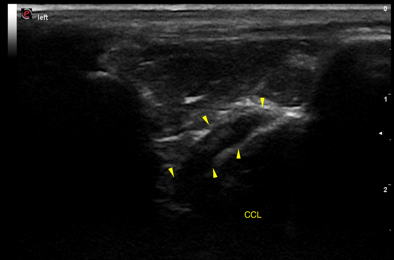
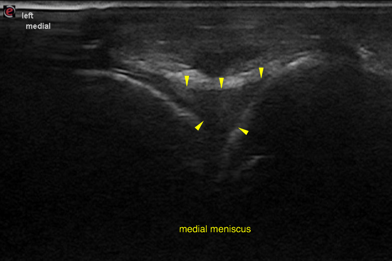
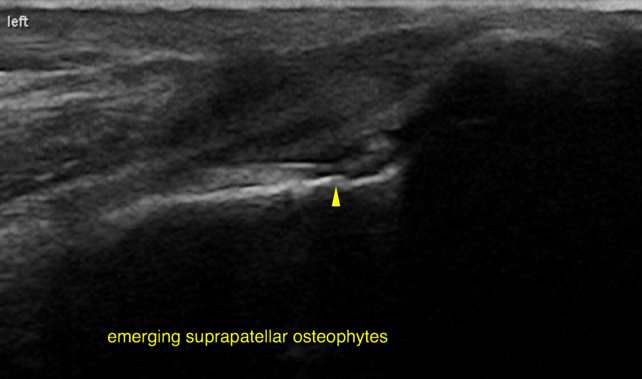
Ultrasound of the stifles, iliopsoas and sartorius muscles – Stifles: Both stifles present intact medial menisci and cranial cruciate ligaments. Joint effusion is not noted. The cartilage layers in the region of the femoral trochlea and the femoral condyles is anechoic, even in thickness and smoothly outlined. The only abnormality noted are very small emerging osteophytes at the proximal aspect of the left femoral trochlea which is likely to be age related and incidental at this stage.
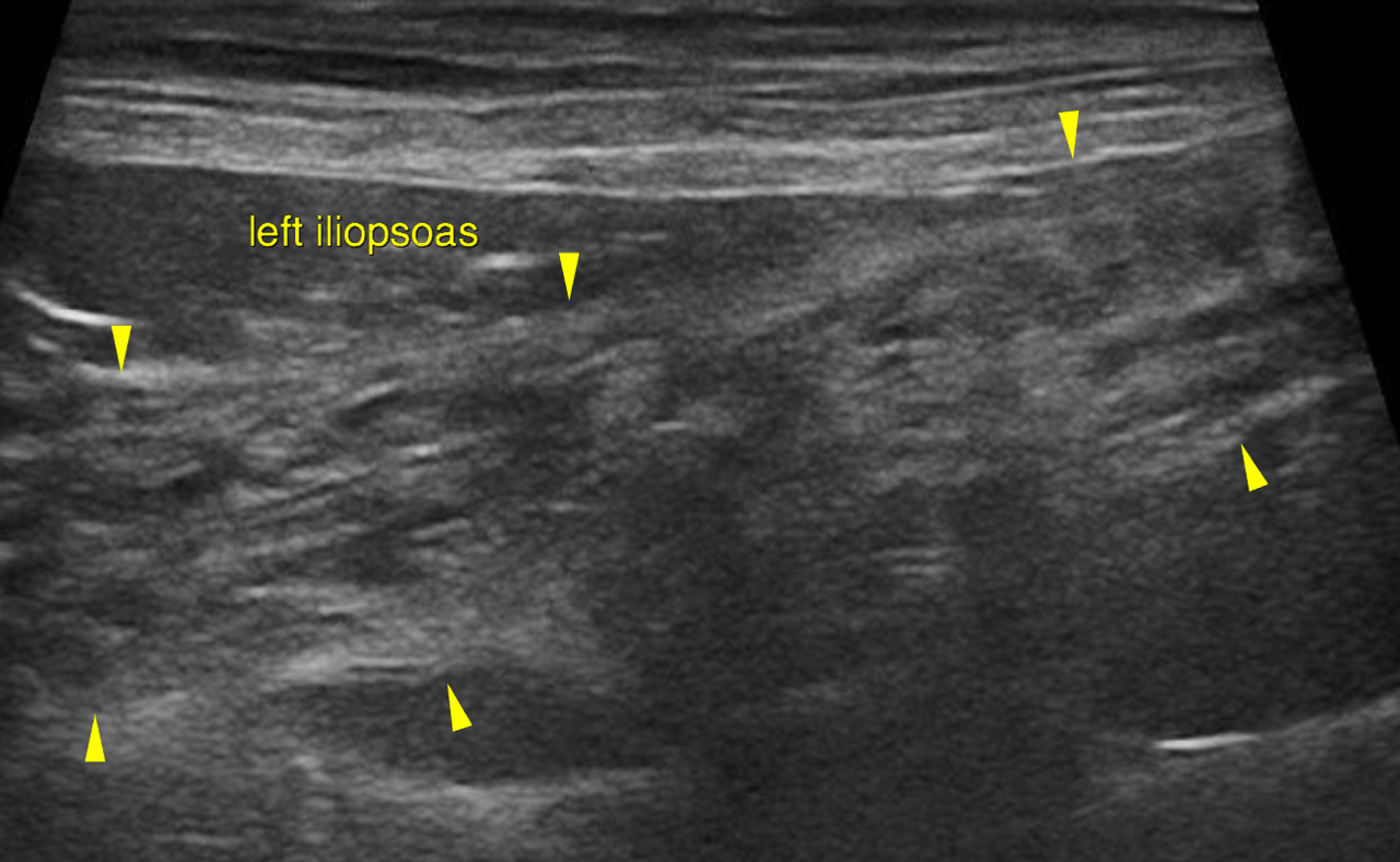
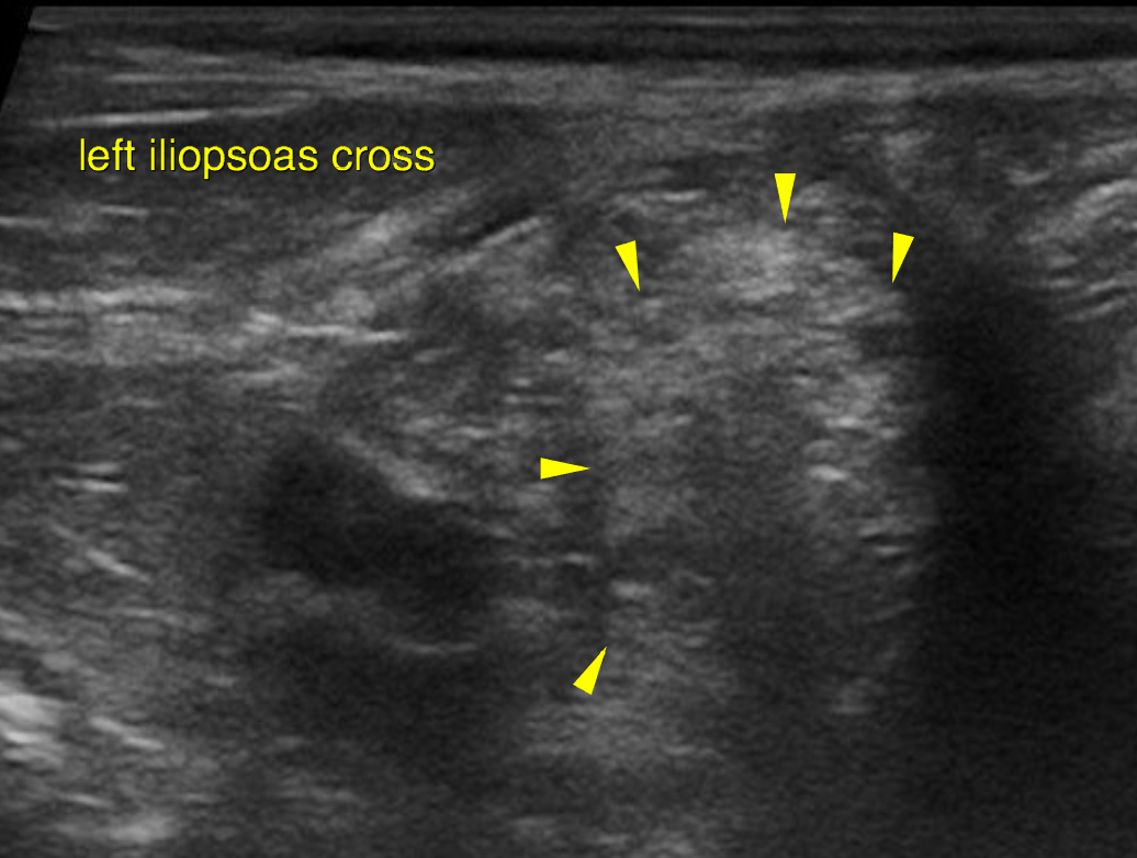
Iliopsoas: The left and right iliopsoas muscle both reveal moderate generalized heterogeneity with an overall increase in echogenicity and mild loss of the regular fiber
pattern.
Multiple small foci causing mild acoustic shadowing are noted throughout the left iliopsoas muscle. No swelling or shape abnormality is evident. The distal insertion of the muscles is not seen.
Sartorius: The left and right sartorius muscle both reveal mild generalized heterogeneity with an overall increase in echogenicity. The proximal and distal insertion of the muscles is not seen.
The degree of echoarchitectural changes is most pronounced for the left iliopsoas muscle and is compatible with emerging multifocal fibrosis.
Conservative treatment with rest, physical therapy and muscular reconditioning (deep heat ultrasound or laser, massages, unforceful stretching) may be attempted. Other orthopedic or neurologic problems – including gastrocnemius musculotendinopathy (high incidence in working herding dogs) – should be ruled out.
Surgical treatment may be considered in case the patient is unresponsive to conservative treatment. Ideally an MRI should be performed to confirm the tentative diagnosis especially when surgical measures are considered.