This 5 year old Terrier cross dog presented for left hind lameness.
This 5 year old Terrier cross dog presented for left hind lameness.
This 5 year old Terrier cross dog presented for left hind lameness.
This 5 year old Terrier cross dog presented for left hind lameness.
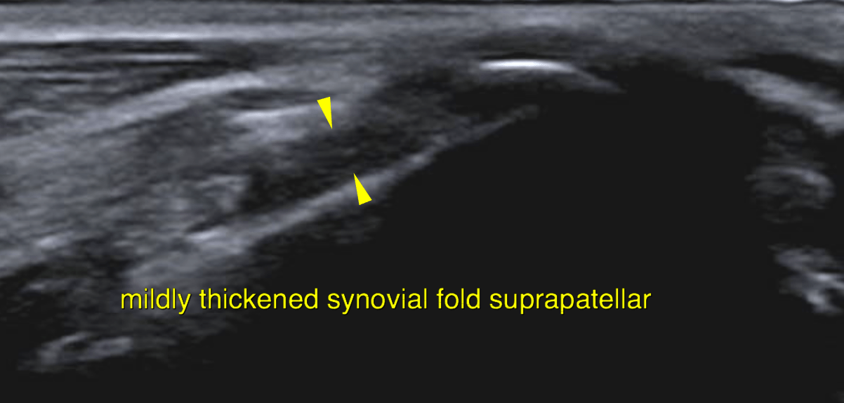
Ultrasound of the right stifle – Both the supra- and infrapatellar recess of the right stifle joint reveal mild synovial
thickening.
The cartilage layers as appreciated onto the femoral trochlea and the femoral condyles
are even and smooth and anechoic as far as seen.
Small emerging osteophytes are seen at the femoral trochlea/patella sulcus.
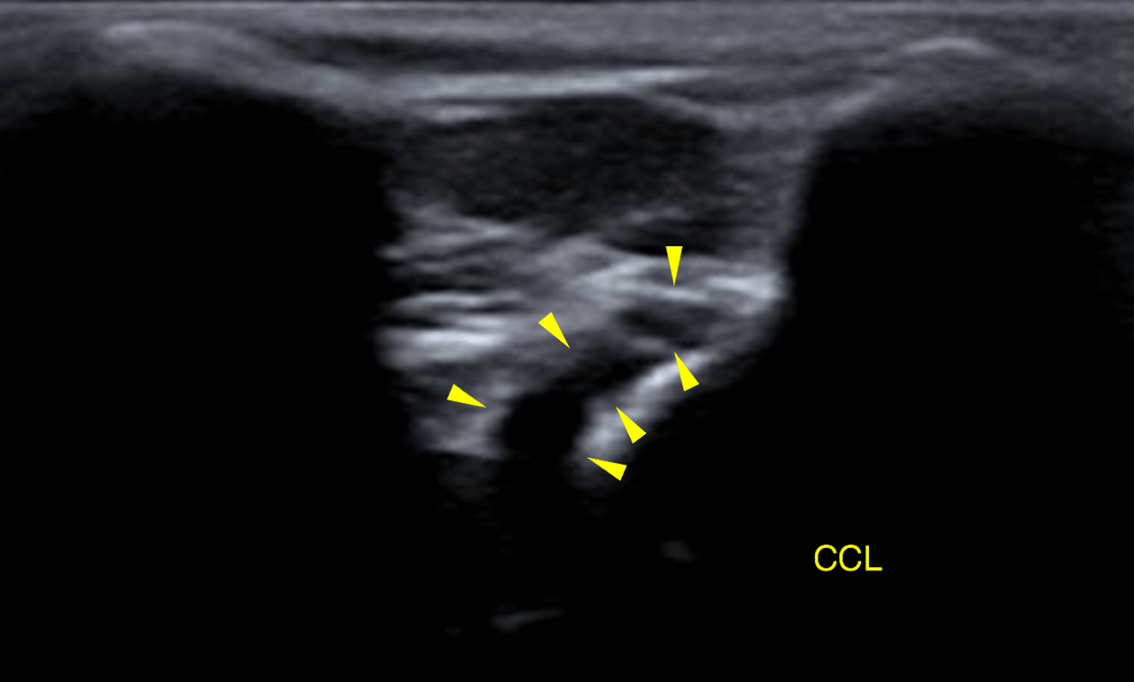
The cranial cruciate ligament (CCL) is continuous and hypoechoic with slightly
undulating contour. There is no periligamentous effusion.
The infrapatellar fat body presents mild heterogeneity with a mild amount of echogenic
tissue in the region oft he CCL as found commonly in chronic degenerative joint
disease (DJD).
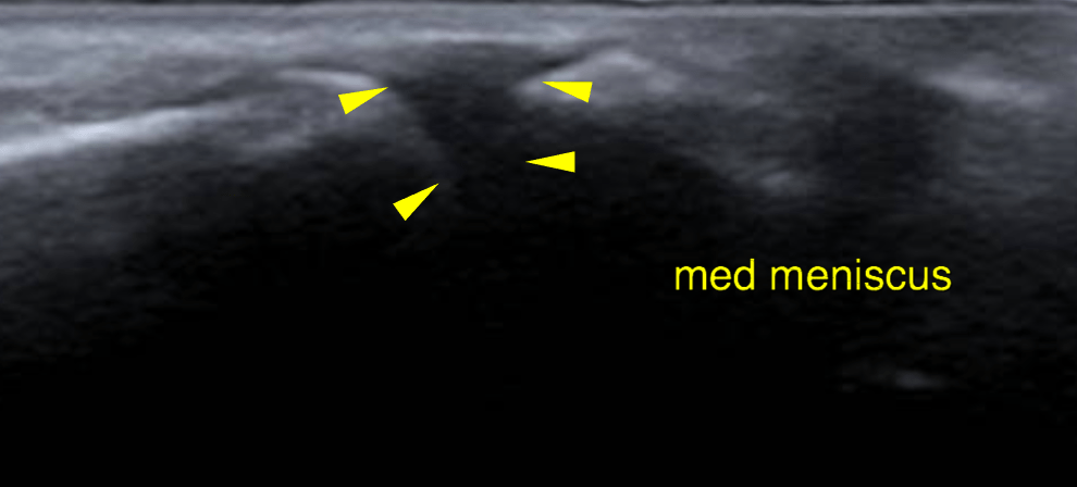
The medial meniscus is well visible. It is in situ with even surface and uniform
echogenicity.
There is no evidence of rupture or bimechanical failure of the CCL. Only mild signs of degeneration are noted.
The medial meniscus is intact.