Progressive transitional cell carcinoma at the cystourethral junction in a 13 year old FS Pit Bull Terrier mix

History

A 13-year-old spayed female Pit Bull Terrier mix dog with history of confirmed transitional cell carcinoma of the cystourethral junction presented for a lack of improvement in clinical signs after antibiotics. The dog had previously been diagnosed with hypothyroidism. Past urinalysis revealed hyposthenuria, hematuria, and pyuria. Recheck urinalysis results were similar to previous results despite antibiotics. The patient was treated with Deramaxx. Several months later, the patient presented for follow-up diagnostics. Urinalysis showed ongoing hematuria and pyuria.

A 13-year-old spayed female Pit Bull Terrier mix dog with history of confirmed transitional cell carcinoma of the cystourethral junction presented for a lack of improvement in clinical signs after antibiotics. The dog had previously been diagnosed with hypothyroidism. Past urinalysis revealed hyposthenuria, hematuria, and pyuria. Recheck urinalysis results were similar to previous results despite antibiotics. The patient was treated with Deramaxx. Several months later, the patient presented for follow-up diagnostics. Urinalysis showed ongoing hematuria and pyuria. CBC was within normal limits, whereas blood chemistry revealed mildly elevated BUN, normal creatinine, and hypercholesterolemia. The patient was treated with Clavamox and famotidine. On recheck urinalysis one month later, 3+ proteinuria, 3+ hematuria, and leukocyturia were still evident.

Clinical Differential Diagnosis

Progressive bladder neoplasia, resistant UTI, uroliths, renal hemorrhage (secondary to neoplasia, uroliths, hypertension, idiopathic etiology), early chronic kidney disease.

DX

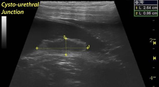
Cystourethral junction mass (transitional cell carcinoma) extending cranially.

Sampling

None taken.

Sonographic Differential Diagnosis

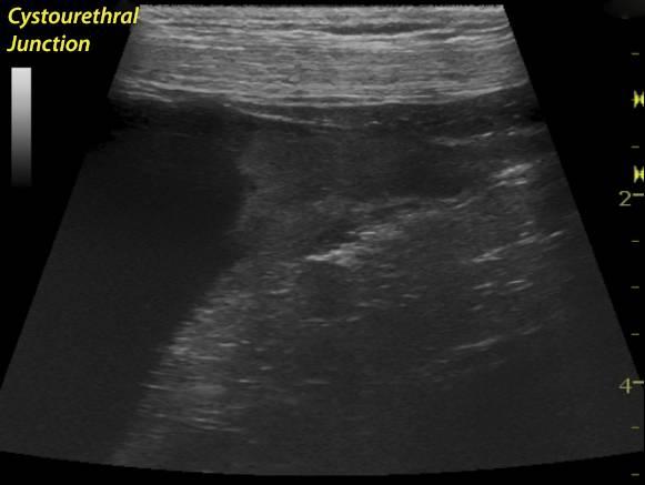
Progressed transitional cell carcinoma of the cystourethral junction, now extending cranially into bladder.

Image Interpretation

The abdomen in this patient presented significantly increasing mass size, measuring 5.13 x 1.79cm in the cystourethral junction and expanding cranially and dorsally along the bladder wall. No evidence of metastatic disease was noted. The urethra was largely normal. No overt obstructive neoplasia was noted within the urethra. No evidence of iliac lymphadenopathy was noted.

Outcome

A repeat of the UGELAB procedure, three view chest radiographs, recheck blood work, and recheck urinalysis were all recommended. The patient was euthanized 10 months after the first UGELAB was performed.

Video

Patient Information

Patient Name : Lucky M
Age : 13 Years
Gender : Female, Spayed
Species : Canine
Book : yes
Liz Wuz Here : Yes
Status : Complete
Code : 06_00016

Blood Chemistry

  • BUN high
  • Cholesterol, High

History

  • Hypothyroidism
  • TCC
  • UTI

Urinalysi

  • Blood Present
  • Protein Present
  • Specific Gravity Low
  • WBCs Present

Images

luckymartellatcccujunction1luckymartellapostugelabtcc1

Skip to content