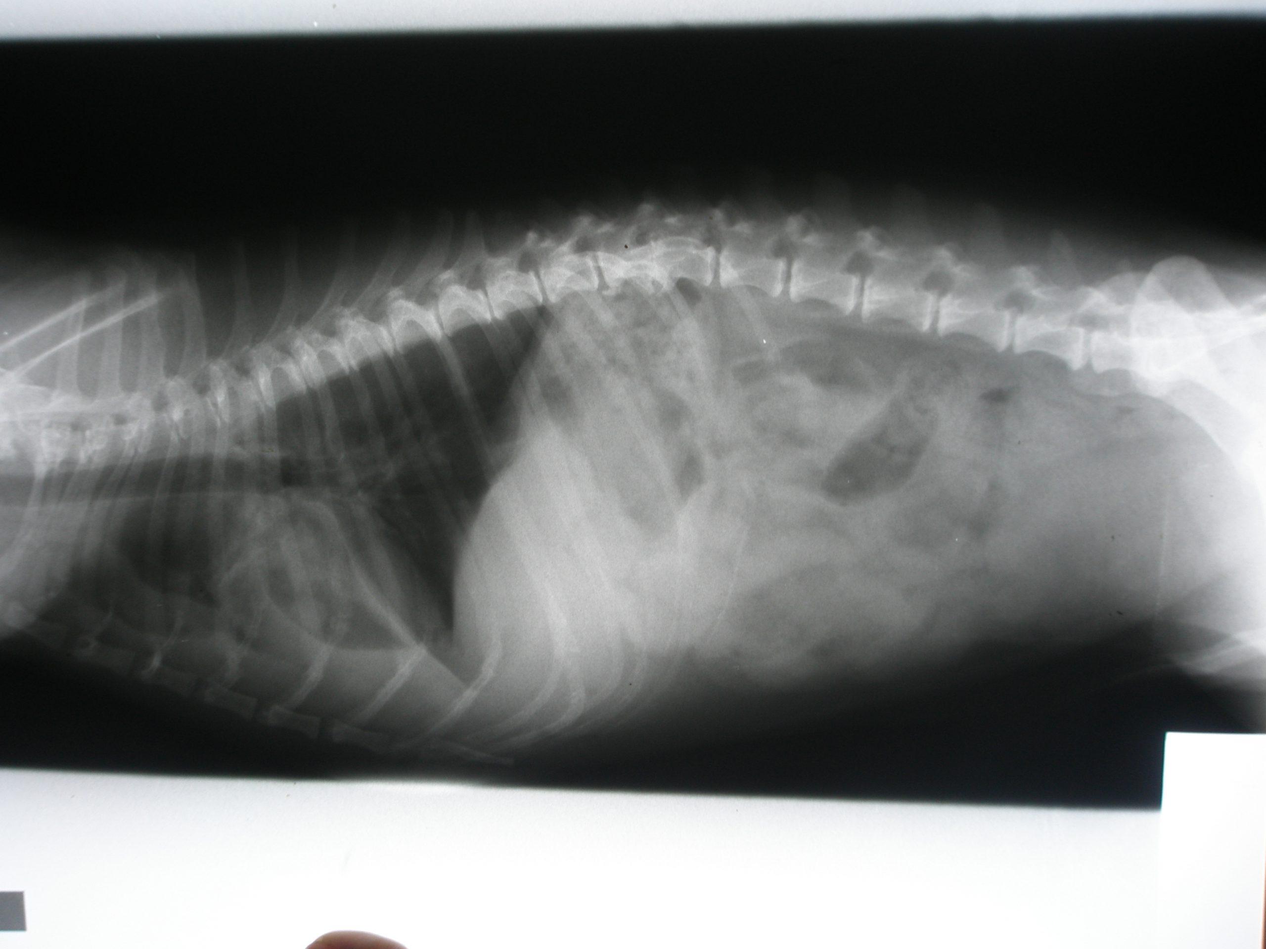
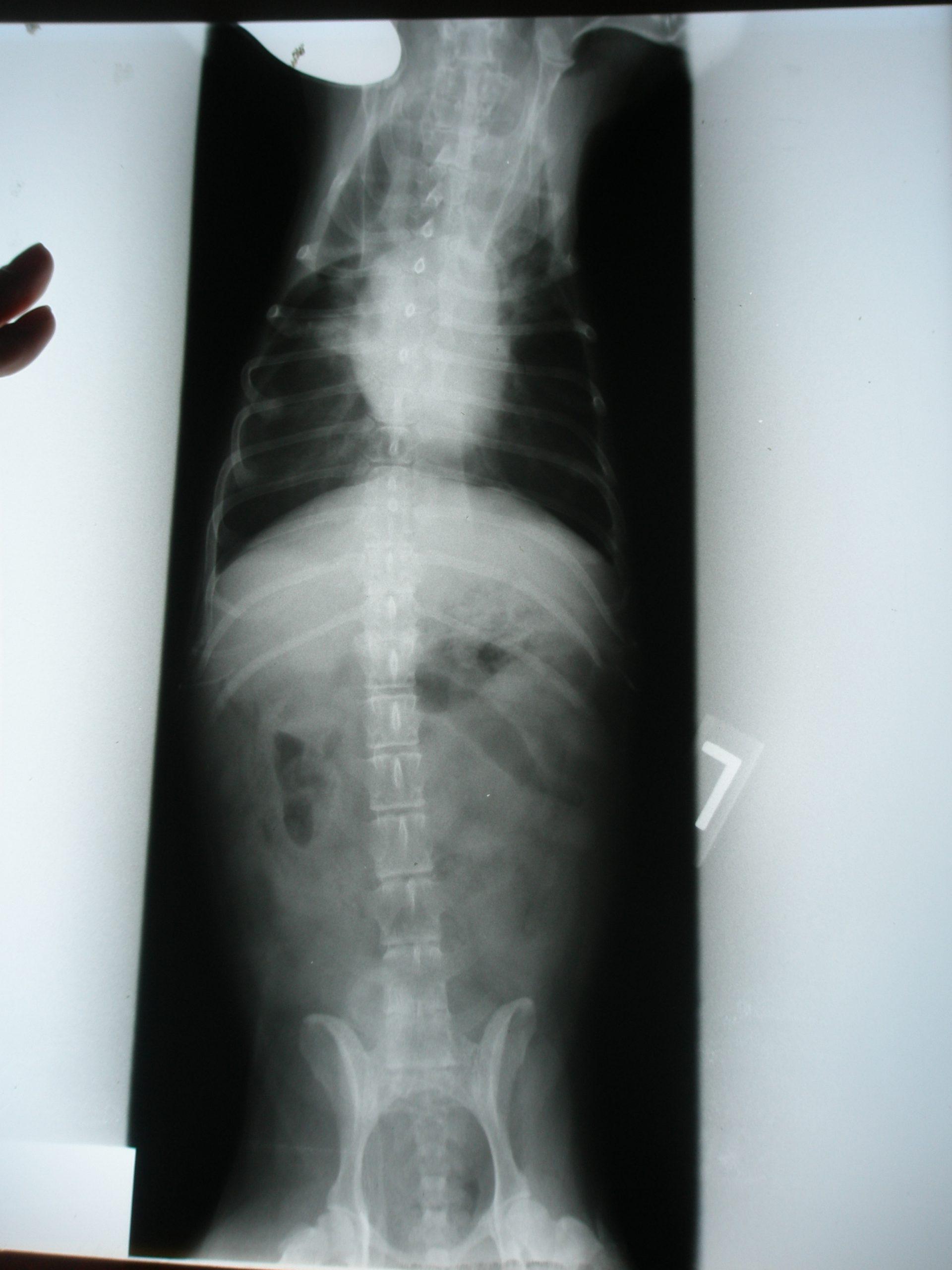
A 19-year-old canine, NM Chihuahua was presented for evaluation of pleural effusion and an abdominal mass. Abnormalities on serum biochemistry were azotemia and elevated cholesterol and triglycerides. Thoracic radiographs revealed pleural effusion and normal cardiac silhouette.
A 19-year-old canine, NM Chihuahua was presented for evaluation of pleural effusion and an abdominal mass. Abnormalities on serum biochemistry were azotemia and elevated cholesterol and triglycerides. Thoracic radiographs revealed pleural effusion and normal cardiac silhouette.
Mass – neoplasia, granuloma, abscess, hydronephrosis, splenic torsion. Pleural effusion – transudate, modified transudate (neoplasia, lung lobe torsion, heart disease), septic exudate, non-septic exudate (blood, chyle).
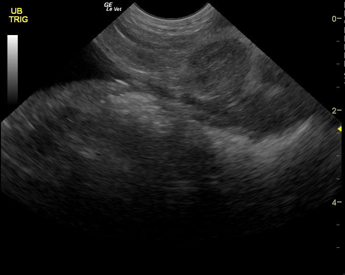
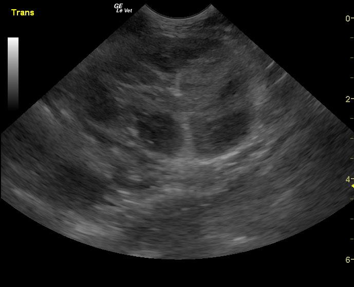
Prostatic mass likely carcinoma, sarcoma less likely.
Prostatic mass with mineralization suggestive for carcinoma. Lymph node metastasis is most likely the cause of sublumbar mass. Normal echocardiogram (not shown) with undefined pleural effusion. Hemorrhagic pleural effusion is a significant concern in this patient; it is likely the abdominal mass has metastasized to the lungs causing necrosis of vasculature and secondary hemorrhage. Prognosis is extremely guarded to poor.