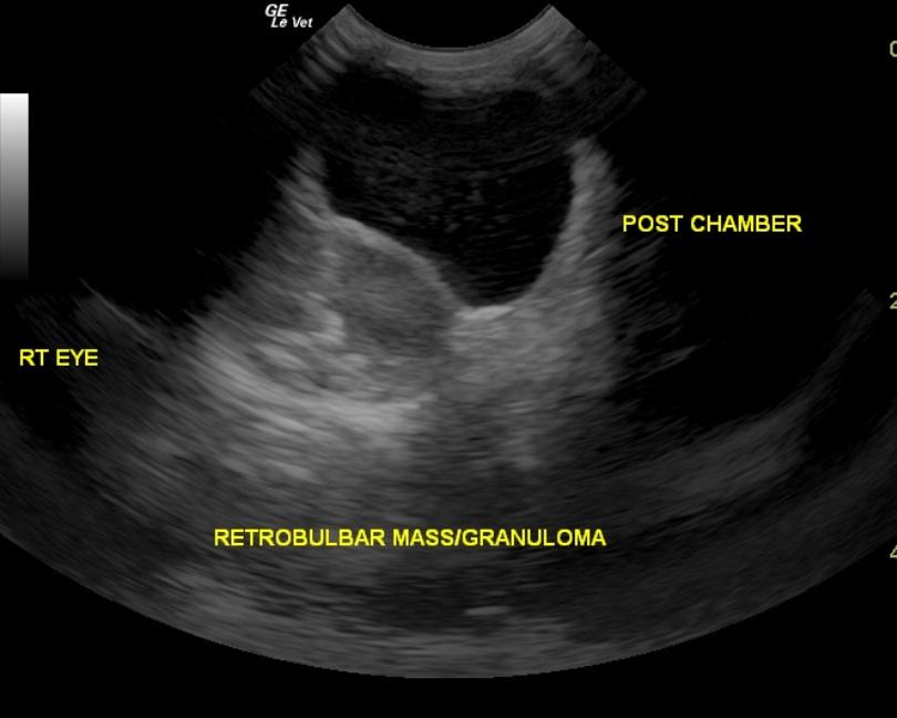
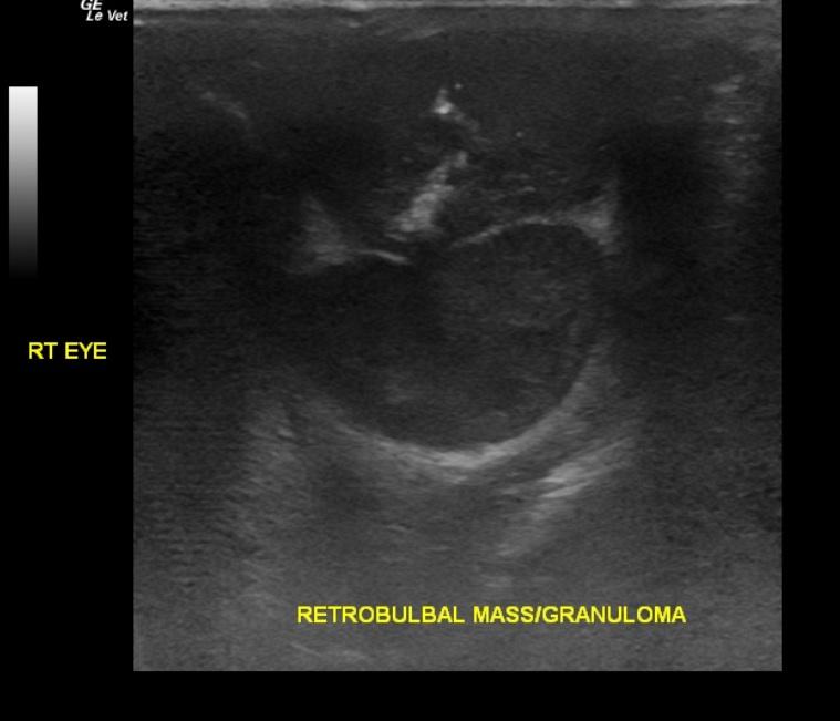
A 12-year-old MN Cocker Spaniel presented for recheck exam after finishing a course of antibiotics for a protruding right eye and a mass behind the eye. The dog had prior medical history of cataracts in both eyes, perianal tumor (removed), and pancreatitis (resolved). Physical exam found the intraocular pressure in the right eye had somewhat improved from last visit, and protrusion of third eyelid had decreased. The dog otherwise appeared healthy.
A 12-year-old MN Cocker Spaniel presented for recheck exam after finishing a course of antibiotics for a protruding right eye and a mass behind the eye. The dog had prior medical history of cataracts in both eyes, perianal tumor (removed), and pancreatitis (resolved). Physical exam found the intraocular pressure in the right eye had somewhat improved from last visit, and protrusion of third eyelid had decreased. The dog otherwise appeared healthy.
Retrobulbar carcinoma or adenocarcinoma diagnosed by FNA in a 12 year old MN Cocker Spaniel
History
Comments
No images of left eye and retrobulbar space are available.
Clinical Differential Diagnosis
Retrobulbar neoplasia, retrobulbar abscess, tooth root abscess
DX
Sampling
US-guided FNA. Cytologic results from aspirate of retrobulbar mass was suggestive of carcinoma or adenocarcinoma with mild, mixed inflammation.
Sonographic Differential Diagnosis
Left eye: chronic inflammatory disease with posterior chamber debris
Right eye: Right retrobulbar mass or organized abscess with periocular tissue inflammation with posterior chamber debris
Image Interpretation
The left eye in this patient presented echogenic debris in the posterior chamber. The retina appeared to be intact however the retrobulbar space and periocular tissues appeared echogenic and irregular. Chronic inflammatory disease is suspected. No overt masses or retrobulbar abscesses were noted of the left eye. The right eye presented a medial retrobulbar abscess or mass. The length of the globe appeared to be wnl. The retina appeared to be intact however the tissue was thickened. The mass was sampled by 25 gauge fine needle aspirate. Fluid was not obtained. Cytology was performed. Granulomatous lesion or neoplastic lesion is suspected. More significant periocular tissue inflammation was noted as well as posterior chamber debris.
Outcome
Patient was discharged with antibiotics and Nsaids, and referral to an ophthalmologist was recommended. The dog was given a very guarded prognosis. At follow-up exam, after antibiotics were finished, the eye was showing little to no improvement; referral to ophthalmologist was recommended again. The next day, the patient presented for pain and scleral hemorrhage and received a steroid injection. Consultation with a specialist was again strongly recommended. Less than 2 weeks later, patient presented back for increased drinking. Exam found third eyelid elevated with mild conjunctivitis. In-house urinalysis was normal. At recheck exam 2 months later, the retrobulbar mass was doubled in size. Owner was to consider removal if dog could no longer close his eye.


