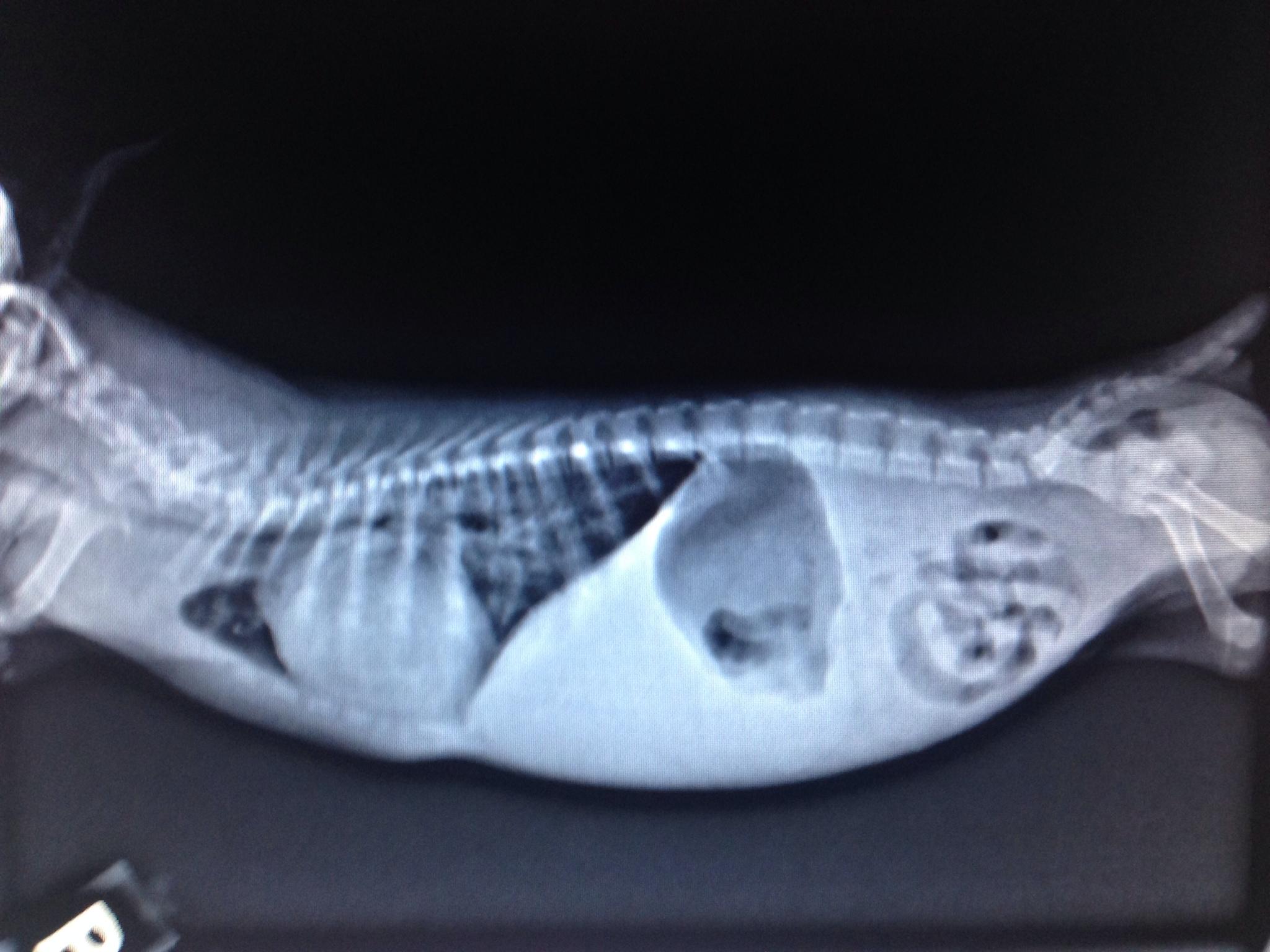
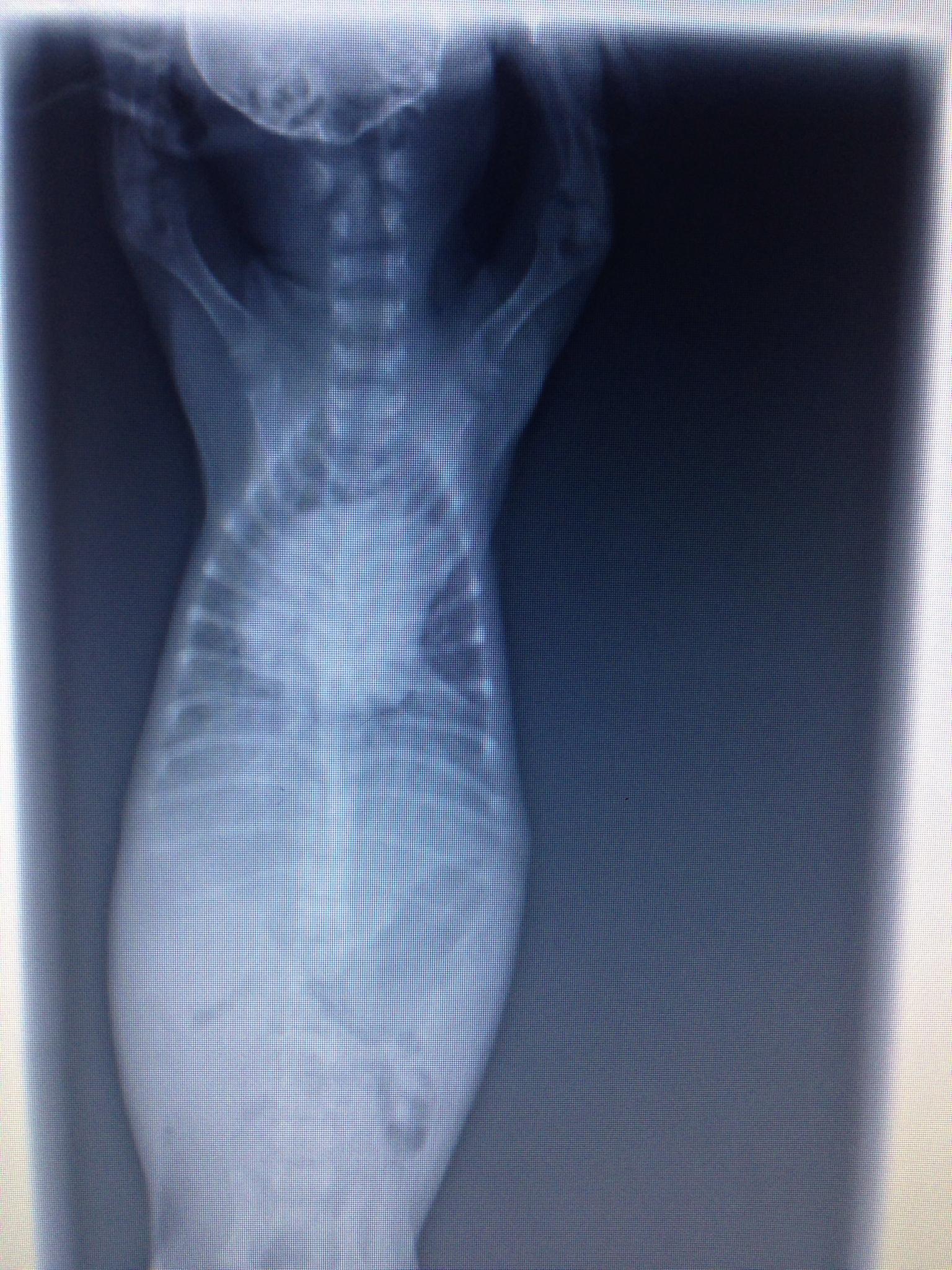
An 8-week-old female Silky Terrier was presented for evaluation of dyspnea and diarrhea. No murmur was auscultated on physical examination. Parvo snap test was negative and hematocrit was 28%. Survey radiographs show right-sided cardiomegaly, focal area of pulmonary consolidation, and an enlarged PA.
An 8-week-old female Silky Terrier was presented for evaluation of dyspnea and diarrhea. No murmur was auscultated on physical examination. Parvo snap test was negative and hematocrit was 28%. Survey radiographs show right-sided cardiomegaly, focal area of pulmonary consolidation, and an enlarged PA.
Lung – pneumonia, edema, atelectasis, hemorrhage, abscess, granuloma
Cardiomegaly – tricuspid dysplasia, atrial/septal defect, pulmonic stenosis, Tetralogy of Fallot
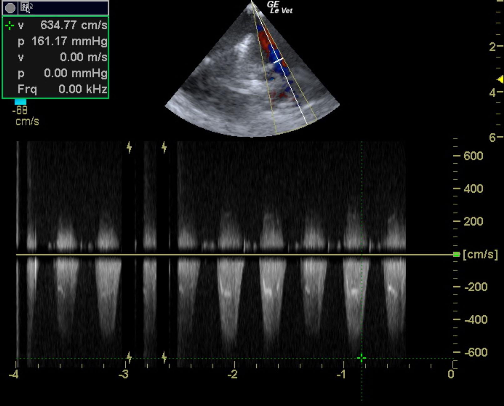
Pulmonary Hypertension – suspected, severe based on presumed TR velocity, degree of LV underloading/pseudohypertrophy and RV concentric hypertrophy
2.) Suggestive for reverse VSD and Eisenmenger’s syndrome
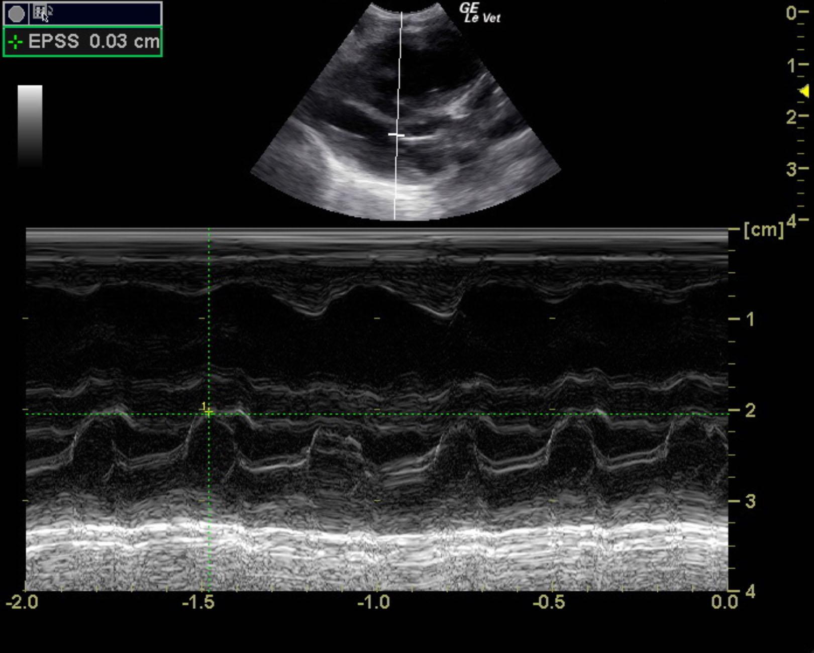
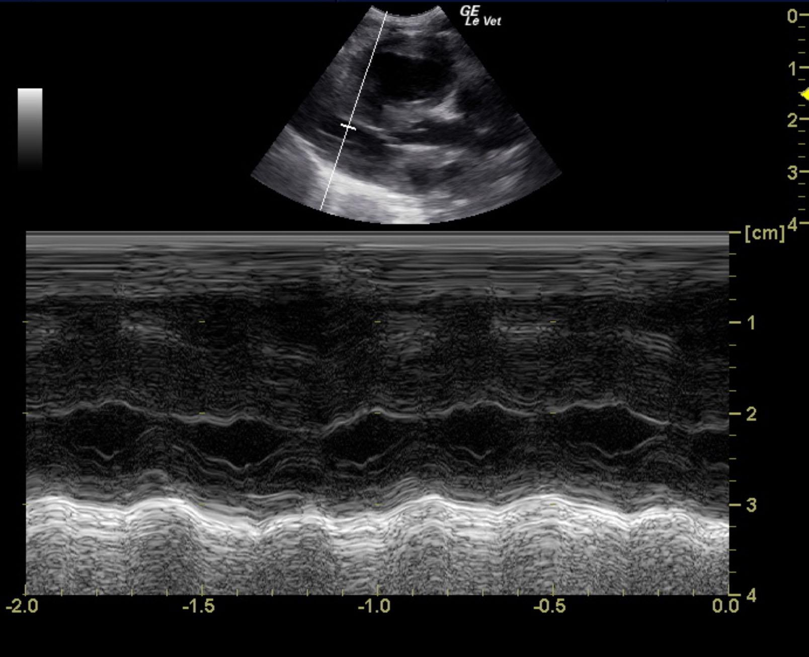
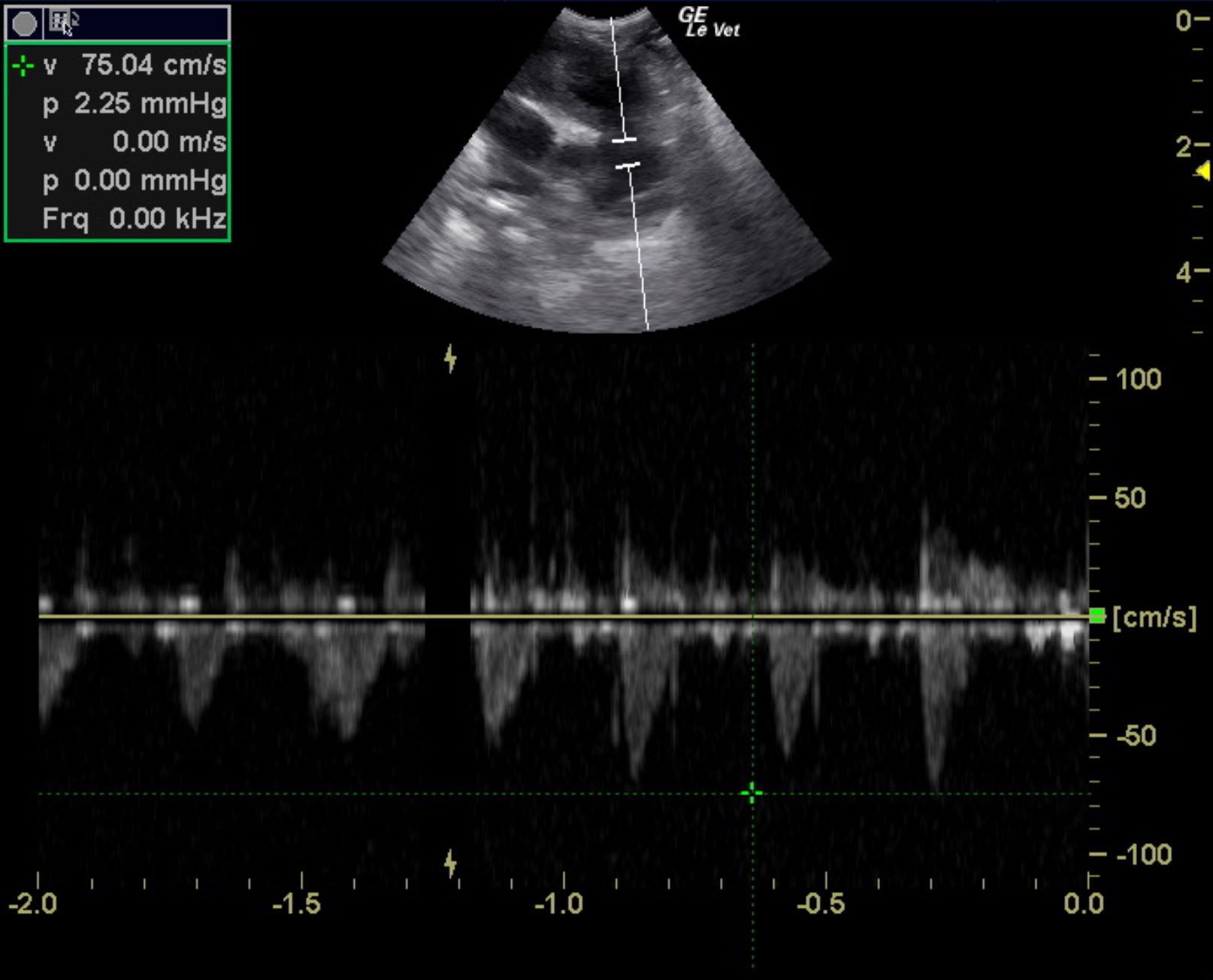
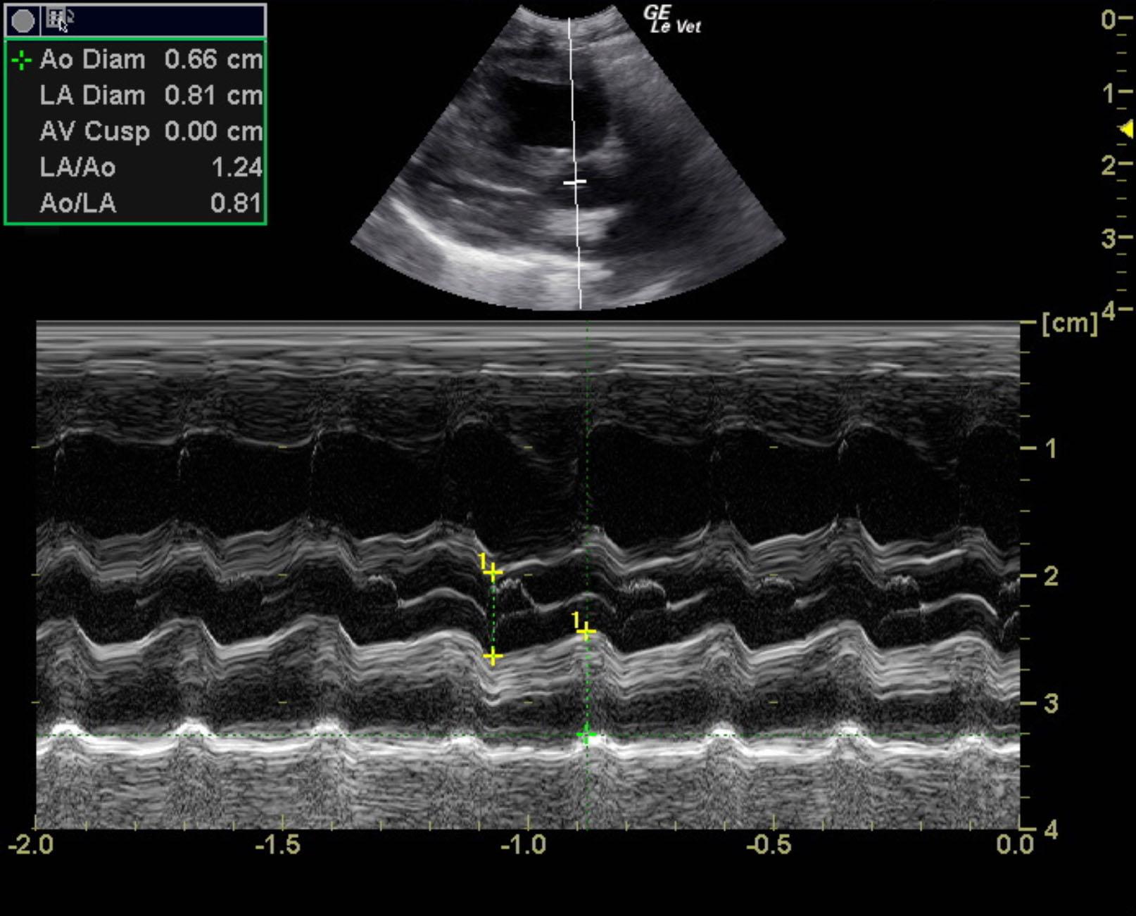
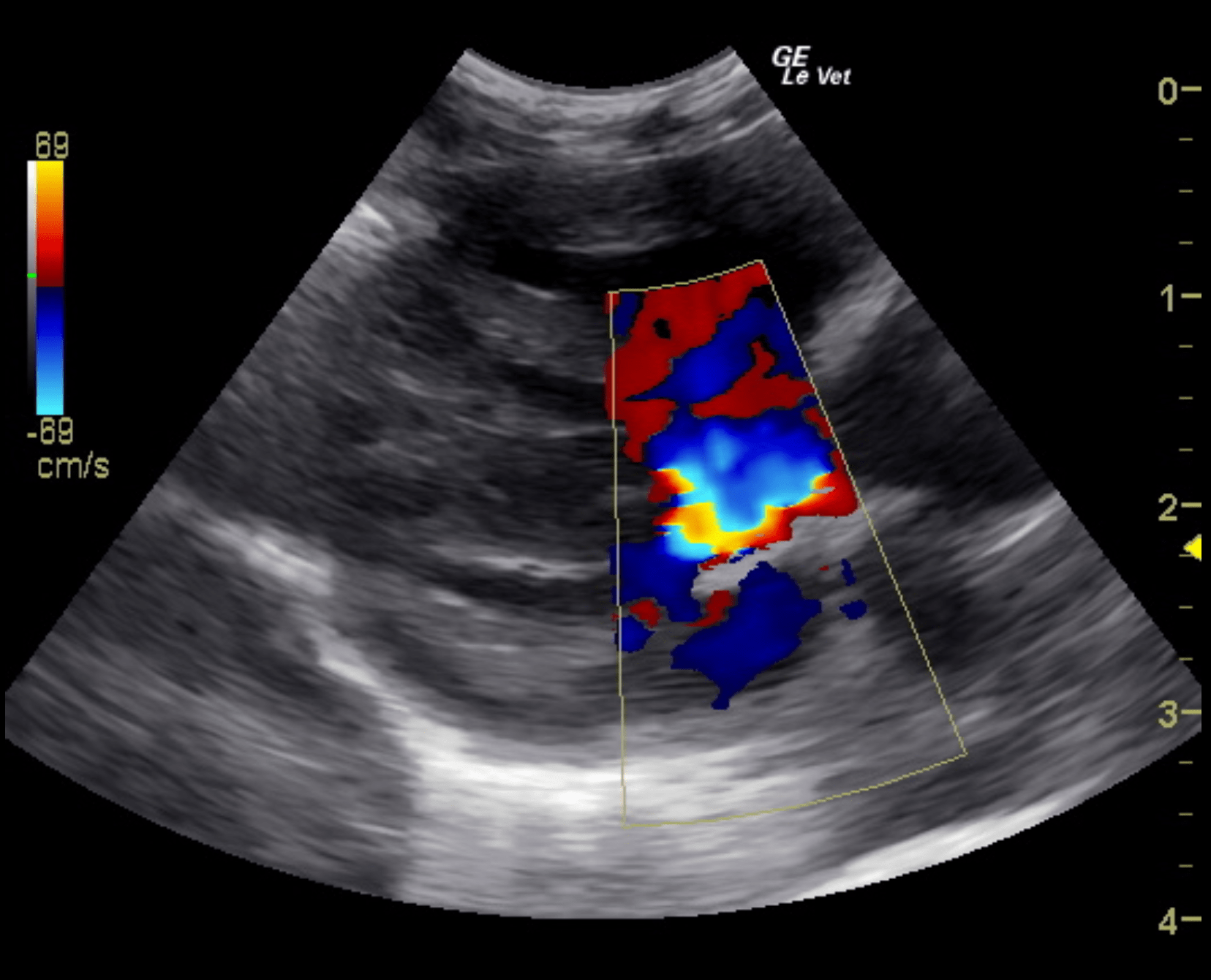
The left ventricular chamber is small with hypertrophied walls, suggestive of volume underload. There is adequate LV function. There is severe dilation and concentric hypertrophy of the RV, subjectively. There is severe RA dilation. The LA is small/normal in size (La:Ao 1.24). The main pulmonary artery is severely enlarged and there appears to gross changes to the branching pulmonary arteries, suggestive of a PDA. There is normal, laminar flow across the PA (0.75m/s). There is high velocity, turbulent systolic flow associated with the septal aspect of the TV that appears to be TR. However, I cannot rule out a VSD. The velocity of flow is high velocity (3.91-6.25m/s). No effusions visualized. LVIDd – 0.91cm FS – ~50%

