This 6-year-old FS Siberian Husky presented for vomiting and anorexia. The patient had been managed for reasonably well controlled diabetes for the previous 6 months. The clinical exam revealed moderate dehydration and abdominal pain. CBC and blood chemistry demonstrated a history of chronic moderately elevated SAP, moderately elevated cholesterol, and moderately elevated triglycerides with slightly elevated lipase. This profile was similar at the time of the clinical presentation with strong evidence of a urinary tract infection on the urinalysis with 3+ glucosuria.
This 6-year-old FS Siberian Husky presented for vomiting and anorexia. The patient had been managed for reasonably well controlled diabetes for the previous 6 months. The clinical exam revealed moderate dehydration and abdominal pain. CBC and blood chemistry demonstrated a history of chronic moderately elevated SAP, moderately elevated cholesterol, and moderately elevated triglycerides with slightly elevated lipase. This profile was similar at the time of the clinical presentation with strong evidence of a urinary tract infection on the urinalysis with 3+ glucosuria. Fructosamine level at that time was also mildly elevated.
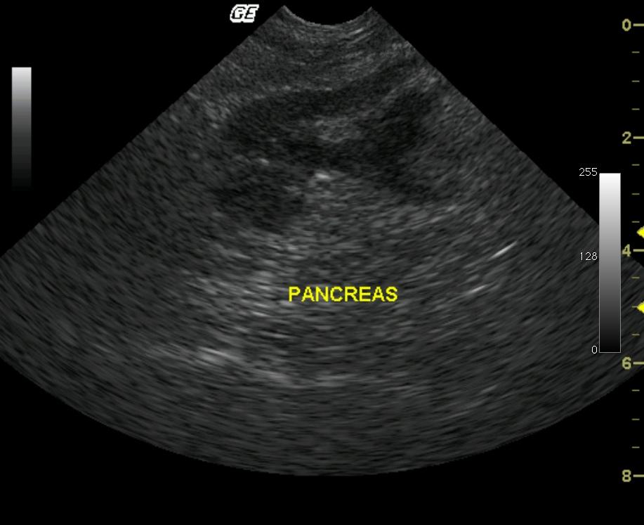
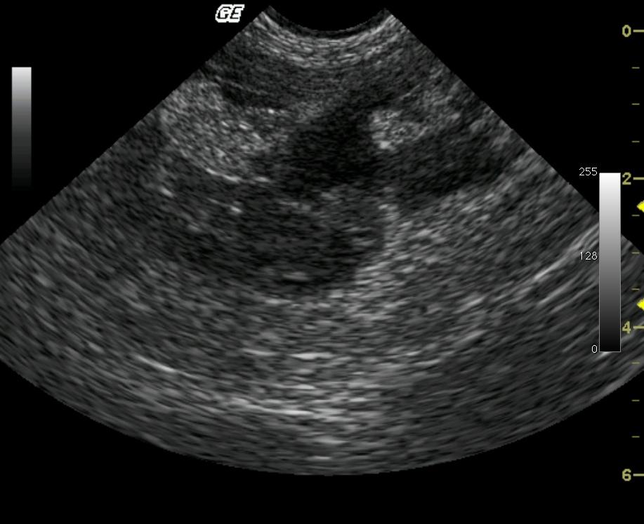
Severe pancreatitis with peripancreatic inflammation in a 6 year old FS Siberian Husky
History
Clinical Differential Diagnosis
Pancreatitis, gastroenteritis, systemic complications to diabetes, pancreatic or other neoplasia.
DX
Sampling
22-gauge US guided FNA of the pancreas revealed low grade mixed suppurative inflammation.
Sonographic Differential Diagnosis
Severe pancreatitis with peripancreatic inflammation. Suspected parenchymal fibrosis suggests acute exacerbation of chronic pancreatitis may have occurred.
Image Interpretation
The imaged portion of the pancreas is enlarged with a markedly hypoechoic parenchyma. The surrounding omentum and mesentery are severely echogenic. Notice the pancreatic margination is irregular. This is likely the result of inflamed omentum overlying the serosal borders. The echogenic pinpoint foci noted within the center of the affected pancreas may represent regional fibrosis, suggesting previous inflammation may have occurred. Color flow Doppler demonstrates persistence of blood flow throughout the affected pancreas.
Outcome
The patient was discharged after 4 days of hospitalization and continued with similar blood values and intermittent episodes over the following 2 years.
Video
Patient Information
Blood Chemistry
- Alkaline Phosphatase (SAP), High
- Cholesterol, High
- Fructosamine, High
- Hypertriglyceridemia
- Lipase, High
Clinical Signs
- Anorexia
- Vomiting
History
- Diabetes, controlled
Exam Finding
- Abdominal Pain
- Dehydration
Urinalysi
- Glucose Present
- WBCs Present
Images