A 12½-year-old neutered male DSH cat with a history of hypertension controlled with amlodipine was presented for evaluation of PU/PD and weight loss. On physical examination, a cranial abdominal mass (suspected kidney) and moderate muscle atrophy was present. Abnormalities on CBC and serum biochemistry included neutrophilia and azotemia. Prior urine analysis had shown proteinuria with a SG of 1.028.
A 12½-year-old neutered male DSH cat with a history of hypertension controlled with amlodipine was presented for evaluation of PU/PD and weight loss. On physical examination, a cranial abdominal mass (suspected kidney) and moderate muscle atrophy was present. Abnormalities on CBC and serum biochemistry included neutrophilia and azotemia. Prior urine analysis had shown proteinuria with a SG of 1.028.
Renal mass: neoplasia, hydronephrosis, pyelonephritis, renolith, granulomatous nephritis.
Bilateral renal lymphoma pattern. Cranial intestinal mass. This is strongly suggestive for multi centric lymphoma primarily involving the kidneys and intestine. Differentials include FIP and aggressive pyelonephritis, but these are much less likely.
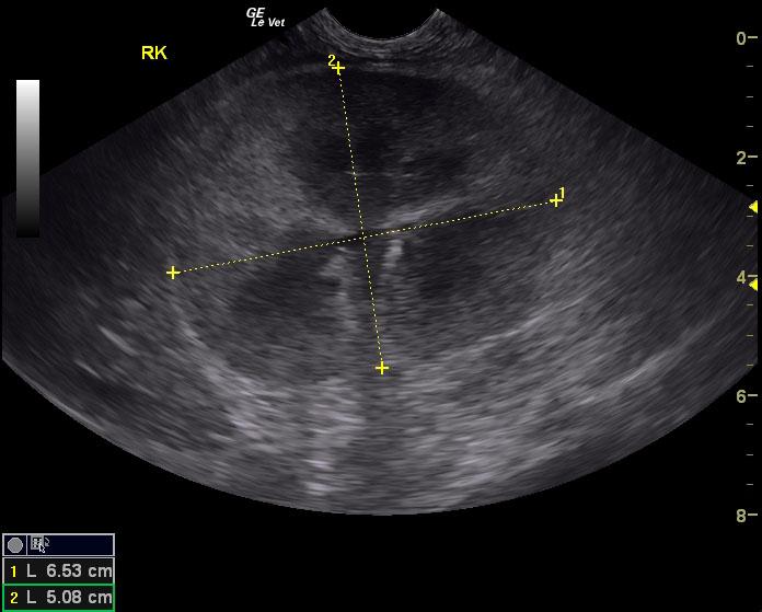
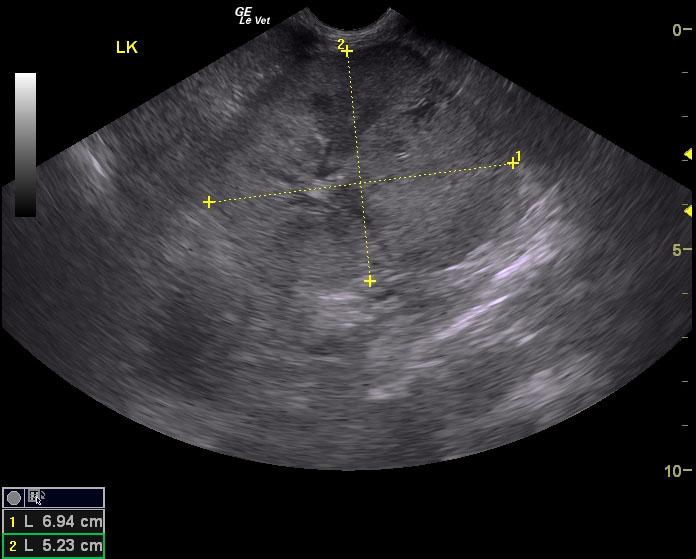
The right kidney was enlarged and measured 6.5 cm with pyelectasia and a pericapsular inflammatory pattern. Hypoechoic parenchyma with multifocal expansive contour was noted. The left kidney presented multiple nodular masses and capsular expansion. Subcapsular capping (the thin hypoechoic line just under the renal capsule) was noted around both kidneys. This is strongly suggestive for lymphoma. A mixed, hypoechoic mass was noted in the small intestine in the cranial abdomen. The mass measured 3.87 x 3.11 cm. Pericapsular inflammatory pattern was noted. The stomach was displaced cranially by the intestinal mass and was empty.